The Influence of Backrest Inclination on Buttock Pressure
- Affiliations
-
- 1Department of Rehabilitation Medicine, Hanyang University College of Medicine, Guri 471-701, Korea. systole@naver.com
- KMID: 2266817
- DOI: http://doi.org/10.5535/arm.2011.35.6.897
Abstract
OBJECTIVE
To assess the effects of backrest inclination of a wheelchair on buttock pressures in spinal cord injured (SCI) patients and normal subjects. METHOD: The participants were 22 healthy subjects and 22 SCI patients. Buttock pressures of the participants were measured by a Tekscan(R) pressure sensing mat and software while they were sitting in a reclining wheelchair. Buttock pressures were recorded for 90degrees, 100degrees, 110degrees, 120degrees and 130degrees seat-to-back angles at the ischial tuberosity (IT) and sacrococcygeal (SC) areas. Recordings were made at each angle over four seconds at a sampling rate of 10 Hz.
RESULTS
The side-to-side buttock pressure differences in the IT area for the SCI patients was significantly greater than for the normal subjects. There was no significant difference between the SCI patients and the normal subjects in the buttock pressure change pattern of the IT area. Significant increases in pressure on the SC area were found as backrest inclination angle was changed to 90degrees, 100degrees and 110degrees in the normal subjects, but no significant differences were found in the SCI patients.
CONCLUSION
Most of the SCI patients have freeform posture in wheelchairs, and this leads to an uneven distribution of buttock pressure. In the SCI patients, the peak pressure in the IT area reduced as the backrest angle was increased, but peak pressure at the SC area remained relatively unchanged. To reduce buttock pressure and prevent pressure ulcers and enhance ulcer healing, it can be helpful for tetraplegic patients, to have wheelchair seat-to-back angles above 120degrees.
Keyword
Figure
-
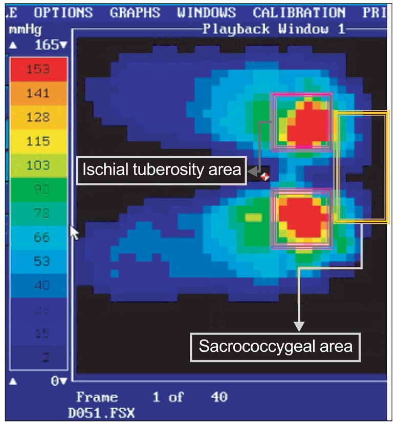
Fig. 1 Buttock pressure distribution in IT-SC areas as displayed on monitor.
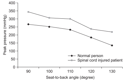
Fig. 2 Changes in the buttock peak pressure of the IT area according to seat-to-back angle in normal subjects and spinal cord injured patients.
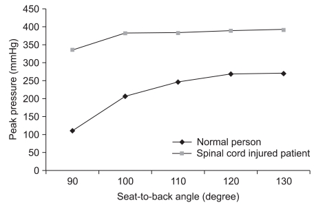
Fig. 3 Changes in the buttock peak pressure of the SC area according to seat-to-back angle in normal subjects and spinal cord injured patients.
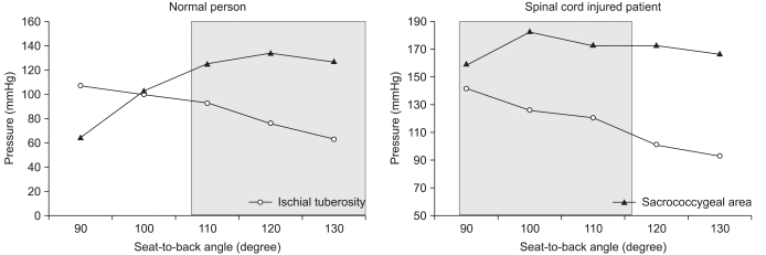
Fig. 4 Patterns of change in average pressure for normal subjects and spinal cord injured patients. The pressure change pattern of the disabled in gray box from 90° seat-to-back angle is similar to that of normal subjects from 105°.
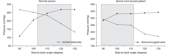
Fig. 5 Paterns of change in peak pressure for normal subjects and spinal cord injured patients. The pressure change pattern of the disabled group in gray box from 90° seat-to-back angle is similar to that of the normal group from 105°
Reference
-
1. Kwon H, Jang IS, Lee JK, Lim P. Clinical observation of the pressure ulcer. J Korean Soc Plast Reconstr Surg. 1996; 23:818–826.2. Uhm KI, Lee YH, Jun KY, Lee YW. Clinical observations of pressure ulcer. J Korean Soc Plast Reconstr Surg. 1980; 7:281–289.3. Dowling AS. Pressure sores-their cause, prevention, and treatment. Md State Med J. 1970; 19:131–134. PMID: 5524138.4. Fuhrer MJ, Garber SL, Rintala DH, Clearman R, Hart KA. Pressure ulcers in community-resident persons with spinal cord injury: prevalence and risk factors. Arch Phys Med Rehabil. 1993; 74:1172–1177. PMID: 8239957.5. Kernozek TW, Lewin JE. Seat interface pressures of individuals with paraplegia: influence of dynamic wheelchair locomotion compared with static seated measurements. Arch Phys Med Rehabil. 1998; 79:313–316. PMID: 9523784.
Article6. Richardson RR, Meyer PR Jr. Prevalence and incidence of pressure ulcers in acute spinal cord injuries. Paraplegia. 1981; 19:235–247. PMID: 7290733.7. Rodriguez GP, Garber SL. Prospective study of pressure ulcer risk in spinal cord injury patients. Paraplegia. 1994; 32:150–158. PMID: 8008417.
Article8. Hackler RH. A 25-year prospective mortality study in the spinal cord injured patient: comparison with long-term living paraplegic. J Urol. 1977; 117:486–488. PMID: 850323.9. Staas WE Jr, Cioschi HM. Pressure sores-a multifaceted approach to prevention and treatment. West J Med. 1991; 154:539–544. PMID: 1830985.10. Pressure sores-a ticking time-bomb. Intensive Crit Care Nurs. 1995; 11:44–48. PMID: 7711400.11. Edberg EL, Cerny K, Stauffer ES. Prevention and treatment of pressure sores. Phys Ther. 1973; 53:246–252. PMID: 4685097.
Article12. Kosiak M. Etiology of decubitus ulcers. Arch Phys Med Rehabil. 1961; 42:19–29. PMID: 13753341.13. Byrne DW, Salzberg CA. Major risk factors for pressure ulcers in the spinal cord disabled: a literature review. Spinal Cord. 1996; 34:255–263. PMID: 8963971.
Article14. Linder-Ganz E, Gefen A. Mechanical compression-induced pressure sores in rat hindlimb: muscle stiffness, histology, and computational models. J Appl Physiol. 2004; 96:2034–2049. PMID: 14766784.
Article15. Allen V, Ryan DW, Lomax N, Murray A. Accuracy of interface pressure measurement systems. J Biomed Eng. 1993; 15:344–348. PMID: 8361161.
Article16. Castro MJ, Apple DF Jr, Staron RS, Campos GE, Dudley GA. Influence of complete spinal cord injury on skeletal muscle within 6 mo of injury. J Appl Physiol. 1999; 86:350–358. PMID: 9887150.
Article17. Modlesky CM, Bickel CS, Slade JM, Meyer RA, Cureton KJ, Dudley GA. Assessment of skeletal muscle mass in men with spinal cord injury using dual-energy X-ray absorptiometry and magnetic resonance imaging. J Appl Physiol. 2004; 96:561–565. PMID: 14527962.
Article18. Gutierrez EM, Alm M, Hultling C, Saraste H. Measuring seating pressure, area, and asymmetry in persons with spinal cord injury. Eur Spine J. 2004; 13:374–379. PMID: 14639505.
Article19. Aissaoui R, Kauffmann C, Dansereau J, de Guise JA. Analysis of pressure distribution at the body-seat interface in able-bodied and paraplegic subjects using a deformable active contour algorithm. Med Eng Phys. 2001; 23:359–367. PMID: 11551812.
Article20. Drummond D, Breed AL, Narechania R. Relationship of spine deformity and pelvic obliquity on sitting pressure distributions and decubitus ulceration. J Pediatr Orthop. 1985; 5:396–402. PMID: 3894415.
Article21. Henderson JL, Price SH, Brandstater ME, Mandac BR. Efficacy of three measures to relieve pressure in seated persons with spinal cord injury. Arch Phys Med Rehabil. 1994; 75:535–539. PMID: 8185445.
Article22. Koo TK, Mak AF, Lee YL. Posture effect on seating interface biomechanics: comparison between two seating cushions. Arch Phys Med Rehabil. 1996; 77:40–47. PMID: 8554472.
Article23. Rosenthal MJ, Felton RM, Hileman DL, Lee M, Friedman M, Navach JH. A wheelchair cushion designed to redistribute sites of sitting pressure. Arch Phys Med Rehabil. 1996; 77:278–282. PMID: 8600872.
Article24. Lancourt JE, Dickson JH, Carter RE. Paralytic spinal deformity following traumatic spinal-cord injury in children and adolesents. J Bone Joint Surg Am. 1981; 63:47–53. PMID: 7451526.25. Hobson DA, Tooms RE. Seated lumbar/pelvic alignment. A comparison between spinal cord-injured and noninjured groups. Spine. 1992; 17:293–229. PMID: 1566167.26. Andersson GB, Murphy RW, Ortengren R, Nachemson AL. The influence of backrest inclination and lumbar support on lumbar lordosis. Spine. 1979; 4:52–58. PMID: 432716.
Article27. Gardocki RJ, Watkins RG, Williams LA. Measurements of lumbopelvic lordosis using the pelvic radius technique as it correlates with sagittal spinal balance and sacral translation. Spine Journal. 2002; 2:421–429. PMID: 14589266.
Article28. Yarkony GM, Bass LM, Keenan V 3rd, Meyer PR Jr. Contractures complicating spinal cord injury: incidence and comparison between spinal cord centre and general hospital acute care. Paraplegia. 1985; 23:265–271. PMID: 4069736.
Article29. Gajdosik RL, Albert CR, Mitman JJ. Influence of hamstring length on the standing position and flexion range of motion of the pelvic angle, lumbar angle, and thoracic angle. J Orthop Sports Phys Ther. 1994; 20:213–219. PMID: 7987382.
Article30. Sumiya T, Kawamura K, Tokuhiro A, Takechi H, Ogata H. A survey of wheelchair use by paraplegic individuals in Japan. Part 2: Prevalence of pressure sores. Spinal Cord. 1997; 35:595–559. PMID: 9300965.
Article31. Schue RM, Langemo DK. Pressure ulcer prevalence and incidence and a modification of the Braden Scale for a rehabilitation unit. J Wound Ostomy Continence Nurs. 1998; 25:36–43. PMID: 9481286.
Article32. Hanson DS, Langemo D, Olson B, Hunter S, Burd C. Evaluation of pressure ulcer prevalence rates for hospice patients post-implementation of pressure ulcer protocols. Am J Hosp Palliat Care. 1994; 11:14–19. PMID: 7893563.
Article33. Shields RK, Cook TM. Effect of seat angle and lumbar support on seated buttock pressure. Phys Ther. 1988; 68:1682–1686. PMID: 3186795.
Article34. Henderson JL, Price SH, Brandstater ME, Mandac BR. Efficacy of three measures to relieve pressure in seated persons with spinal cord injury. Arch Phys Med Rehabil. 1994; 75:535–539. PMID: 8185445.
Article35. Tam EW, Mak AF, Lam WN, Evans JH, Chow YY. Pelvic movement and interface pressure distribution during manual wheelchair propulsion. Arch Phys Med Rehabil. 2003; 84:1466–1472. PMID: 14586913.36. Bush CA. Study of pressures on skin under ischial tuberosities and thighs during sitting. Arch Phys Med Rehabil. 1969; 50:207–213. PMID: 5779442.
- Full Text Links
-
- Actions
-
Cited
- CITED
-
- Close
- Share
-
- Similar articles
-
- Effects of Backrest Position on Central Venous Pressure and Intracranial Pressure in Brain Surgery Patients
- Reduction Method of Anterior Shoulder Dislocation: A New Method
- The Effect of Chair Backrest on Respiratory Function in Prolonged Sitting Position
- Comparison of the Symmetry of Buttock Pressure during Simulated Driving between Heathy Adults and Patients with Stroke
- The Effects of Pressure Relief Methods at Wheelchair Seated Spinal Cord Injured Patients
