Fingernail Onychomycosis Due to Aspergillus niger
- Affiliations
-
- 1Department of Dermatology, College of Medicine, Dongguk University, Gyeongju, Korea. smg@dongguk.ac.kr
- 2Department of Laboratory Medicine, College of Medicine, Dongguk University, Gyeongju, Korea.
- 3Department of Dermatology, College of Medicine, Yeungnam University, Daegu, Korea.
- KMID: 2266043
- DOI: http://doi.org/10.5021/ad.2012.24.4.459
Abstract
- Onychomycosis is usually caused by dermatophytes, but some species of nondermatophytic molds and yeasts are also associated with nail invasion. Aspergillus niger is a nondermatophytic mold which exists as an opportunistic filamentous fungus in all environments. Here, we report a case of onychomycosis caused by A. niger in a 66-year-old female. The patient presented with a black discoloration and a milky white base and onycholysis on the proximal portion of the right thumb nail. Direct microscopic examination of scrapings after potassium hydroxide (KOH) preparation revealed dichotomous septate hyphae. Repeated cultures on Sabouraud's dextrose agar (SDA) without cycloheximide produced the same black velvety colonies. No colony growth occurred on SDA with cycloheximide slants. Biseriate phialides covering the entire vesicle with radiate conidial heads were observed on the slide culture. The DNA sequence of the internal transcribed spacer region of the clinical sample was a 100% match to that of A. niger strain ATCC 16888 (GenBank accession number AY373852). A. niger was confirmed by KOH mount, colony identification, light microscopic morphology, and DNA sequence analysis. The patient was treated orally with 250 mg terbinafine daily and topical amorolfine 5% nail lacquer for 3 months. As a result, the patient was completely cured clinically and mycologically.
Keyword
MeSH Terms
-
Agar
Aged
Arthrodermataceae
Aspergillus
Aspergillus niger
Base Sequence
Cycloheximide
Female
Fungi
Glucose
Head
Humans
Hydroxides
Hyphae
Lacquer
Light
Morpholines
Nails
Naphthalenes
Niger
Onycholysis
Onychomycosis
Potassium
Potassium Compounds
Sequence Analysis, DNA
Sprains and Strains
Thumb
Yeasts
Agar
Cycloheximide
Glucose
Hydroxides
Morpholines
Naphthalenes
Potassium
Potassium Compounds
Figure
-
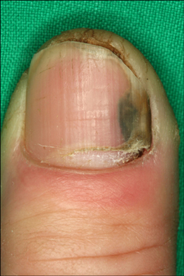
Fig. 1 Black discoloration with milky white base and onycholysis on the proximal portion of the right thumb nail.

Fig. 2 Direct microscopic examination of scraping from a potassium hydroxide (KOH) preparation revealing dichotomous septate hyphae (KOH mount, ×400).

Fig. 3 Multiple, black colonies with velvety surfaces on Sabouraud's dextrose agar slants after 1 week at 25℃.

Fig. 4 A rapidly growing, black colony with a velvety surface on an Sabouraud's dextrose agar plate after 5 days at 25℃.

Fig. 5 Biseriate phialides covering the entire vesicle with radiate conidial heads in a slide culture of Aspergillus niger (lactophenol-cotton blue stain, ×400).

Fig. 6 Alignment of internal transcribed spacer (ITS) sequences showing the ITS sequence from the clinical sample was a 100% match to that of Aspergillus niger strain ATCC 16888 (GenBank accession number AY373852).
Reference
-
1. Verma S, Heffeman MP. Wolff K, Goldsmith LA, Katz SI, Gilchrest BA, Paller AS, Leffell DJ, editors. Superficial fungal infection: dermatophytosis, onychomycosis, tinea nigra, piedra. Fitzpatrick's dermatology in general medicine. 2008. 7th ed. New York: McGraw-Hill;1807–1821.2. Moreno G, Arenas R. Other fungi causing onychomycosis. Clin Dermatol. 2010. 28:160–163.
Article3. Bassiri-Jahromi S, Khaksar AA. Nondermatophytic moulds as a causative agent of onychomycosis in tehran. Indian J Dermatol. 2010. 55:140–143.
Article4. Gianni C, Cerri A, Crosti C. Non-dermatophytic onychomycosis. An understimated entity? A study of 51 cases. Mycoses. 2000. 43:29–33.
Article5. Gupta AK, Ryder JE, Baran R, Summerbell RC. Non-dermatophyte onychomycosis. Dermatol Clin. 2003. 21:257–268.
Article6. Romano C, Gianni C, Difonzo EM. Retrospective study of onychomycosis in Italy: 1985-2000. Mycoses. 2005. 48:42–44.
Article7. Gupta M, Sharma NL, Kanga AK, Mahajan VK, Tegta GR. Onychomycosis: clinico-mycologic study of 130 patients from Himachal Pradesh, India. Indian J Dermatol Venereol Leprol. 2007. 73:389–392.
Article8. Gianni C, Romano C. Clinical and histological aspects of toenail onychomycosis caused by Aspergillus spp.: 34 cases treated with weekly intermittent terbinafine. Dermatology. 2004. 209:104–110.
Article9. Hilmioğlu-Polat S, Metin DY, Inci R, Dereli T, Kilinç I, Tümbay E. Non-dermatophytic molds as agents of onychomycosis in Izmir, Turkey-a prospective study. Mycopathologia. 2005. 160:125–128.
Article10. Lim SW, Suh MK, Ha GY. Clinical features and identification of etiologic agents in onychomycosis (1999-2002). Korean J Dermatol. 2004. 42:53–60.11. Tosti A, Piraccini BM, Lorenzi S. Onychomycosis caused by nondermatophytic molds: clinical features and response to treatment of 59 cases. J Am Acad Dermatol. 2000. 42:217–224.
Article12. Surjushe A, Kamath R, Oberai C, Saple D, Thakre M, Dharmshale S, et al. A clinical and mycological study of onychomycosis in HIV infection. Indian J Dermatol Venereol Leprol. 2007. 73:397–401.
Article13. Tosti A, Piraccini BM. Proximal subungual onychomycosis due to Aspergillus niger: report of two cases. Br J Dermatol. 1998. 139:156–157.
Article14. Summerbell RC, Kane J, Krajden S. Onychomycosis, tinea pedis and tinea manuum caused by non-dermatophytic filamentous fungi. Mycoses. 1989. 32:609–619.
Article15. Rippon JW. Medical mycology: the pathogenic fungi and the pathogenic actinomycetes. 1988. 3rd ed. Philadelphia: WB Saunders;618–650.16. Böhler K, Metze D, Poitschek C, Jurecka W. Cutaneous aspergillosis. Clin Exp Dermatol. 1990. 15:446–450.
Article17. Takahata Y, Hiruma M, Sugita T, Muto M. A case of onychomycosis due to Aspergillus sydowii diagnosed using DNA sequence analysis. Mycoses. 2008. 51:170–173.
Article18. Grover S. Clinico-mycological evaluation of onychomycosis at Bangalore and Jorhat. Indian J Dermatol Venereol Leprol. 2003. 69:284–286.19. De Hoog GS, Guarro J, Gene J, Figueras MJ. Atlas of clinical fungi. 2000. 2nd ed. Utrecht: Centraalbureau voor Schimmelcultures;442–519.20. Suh MK, Ha GY. A clinical and mycological study of otomycosis. Korean J Med Mycol. 1999. 4:15–20.21. English MP. Nails and fungi. Br J Dermatol. 1976. 94:697–701.
Article22. Makimura K, Tamura Y, Mochizuki T, Hasegawa A, Tajiri Y, Hanazawa R, et al. Phylogenetic classification and species identification of dermatophyte strains basedon DNA sequences of nuclear ribosomal internal transcribed spacer 1 regions. J Clin Microbiol. 1999. 37:920–924.
Article23. Kwon-Chung KJ, Bennet JE. Medical mycology. 1992. Philadelphia: Lea & Febiger;201–247.24. Baran R, Hay RJ, Tosti A, Haneke E. A new classification of onychomycosis. Br J Dermatol. 1998. 139:567–571.
Article25. Nolting S, Brautigam M, Weidinger G. Terbinafine in onychomycosis with involvement by non-dermatophytic fungi. Br J Dermatol. 1994. 130:Suppl 43. 16–21.
Article
- Full Text Links
-
- Actions
-
Cited
- CITED
-
- Close
- Share
-
- Similar articles
-
- A Case of Fingernail Onychomycosis Caused by Fusarium proliferatum
- A Case of White Superficial Onychomycosis
- A Case of Primary Cutaneous Infection of the Burn Scar by Aspergillus niger
- A Case of Primary Cutaneous Scar Infection Caused by Aspergillus niger
- A Case of Primary Cutaneous Aspergillosis of the Dorsum of Hand Due to Aspergillus niger Manifested by Edematous Lesion
