Three Cases of 'Morsicatio Labiorum'
- Affiliations
-
- 1Department of Dermatology, Hanyang University College of Medicine, Seoul, Korea.
- 2Department of Dermatology, Jesus Hospital/Presbyterian Medical Center, Jeonju, Korea. cwlee@hanyang.ac.kr
- KMID: 2266042
- DOI: http://doi.org/10.5021/ad.2012.24.4.455
Abstract
- Morsicatio labiorum is a form of tissue alteration caused by self-induced injury, mostly occurring on the lips, and is considered to be a rarely encountered mucocutaneous disorder. Clinically, it is a macerated grey-white patch and plaque of the mucosa caused by external stimuli (self-induced injury) such as habitual biting, chewing, or sucking of the lip. It is often confused with other dermatological disorders involving the oral mucosa, which can lead to a misdiagnosis. We herein report three cases of morsicatio labiorum; two cases were misdiagnosed as exfoliative cheilitis at the time of the first visit.
Keyword
MeSH Terms
Figure
-
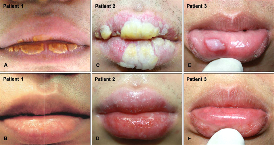
Fig. 1 (A, C) Localized yellow hyperkeratotic plaques on the upper and lower lips in patients 1 & 2. (E) A well-demarcated, white, smooth plaque on the lower lip in patient 3. (B, D, F) The lesions had completely disappeared after the patients retained from their lesion causing habits.
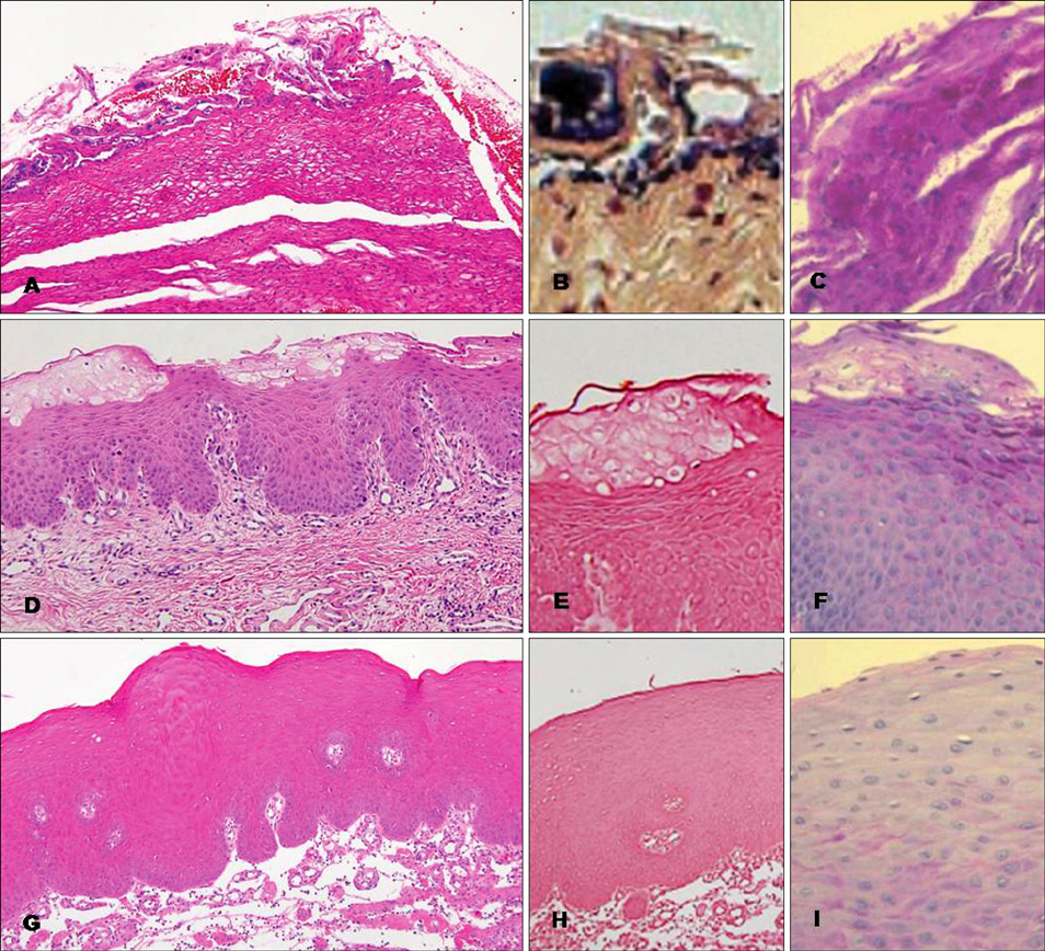
Fig. 2 (A) Histopathologic examination of the underneath mucosal surface of the plaque showed focal hyperkeratosis with basophilic debris on the surface (H&E, ×100). (B) Gram positive (purple-colored) bacterial colonies were found (Gram stain, ×100). (C) The specimen was negative for fungal infection (PAS stain, ×400). (D) The epidermis showed marked acanthosis and necrotic keratinocytes. Beneath the surface, swollen keratinocytes were seen (H&E, ×100). (E) Bacteria colonization was not observed (Gram stain, ×100). (F) The specimen was negative for fungal infection (PAS stain, ×400). (G) The epidermis showed marked acanthosis; however, bacteria colonization was not seen (H&E, ×100). (H) A gram stain was negative (Gram stain, ×100). (I) The specimen was negative for fungal infection (PAS stain, ×400).
Reference
-
1. Glass LF, Maize JC. Morsicatio buccarum et labiorum (excessive cheek and lip biting). Am J Dermatopathol. 1991. 13:271–274.
Article2. Tomás Carmona I, Cameselle Tejeiro J, Diz Dios P, Seoane Lestón J, Castro Ferreiro M, Limeres Posses J. Morsicatio linguarum versus oral hairy leukoplakia. Dermatology. 2000. 201:281–282.
Article3. Scully C, Hegarty A. Burns T, Breathnach S, Cox N, Griffiths C, editors. The oral cavity and lips. Rook's textbook of dermatology. 2010. 8th ed. Massachusetts: Blackwell;69.87–69.88.
Article4. Woo SB, Lin D. Morsicatio mucosae oris--a chronic oral frictional keratosis, not a leukoplakia. J Oral Maxillofac Surg. 2009. 67:140–146.
Article5. Allen AM, Camisa C. Callen JP, Horn TD, Mancini AJ, Salasche SJ, Schaffer JV, Schwarz T, editors. Oral disease. Dermatology. 2008. 2nd ed. Spain: Mosby Elsevier;1044.6. Obermayer ME. Cheekbiting (morsicatio buccarum). Arch Dermatol. 1964. 90:185–190.
Article7. Van Wyk CW, Staz J, Farman AG. The chewing lesion of the cheeks and lips: its features and prevalence among a selected group of adolescents. J Dent. 1977. 5:193–199.
Article
