Ann Dermatol.
2013 Aug;25(3):396-397. 10.5021/ad.2013.25.3.396.
Woolly Hair Nevus Involving Entire Occipital and Temporal Scalp
- Affiliations
-
- 1Department of Dermatology and Institute of Hair and Cosmetic Medicine, Yonsei University Wonju College of Medicine, Wonju, Korea. leewonsoo@yonsei.ac.kr
- KMID: 2265894
- DOI: http://doi.org/10.5021/ad.2013.25.3.396
Abstract
- No abstract available.
Figure
-
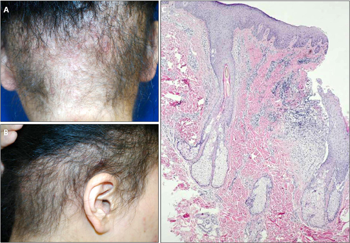
Fig. 1 Well-defined patch of abnormal curly hair over the occipital and lower temporal area. (A) Right 90° side view. (B) Posterior view. (C) Wavy hair follicles with perifollicular infiltration of the inflammatory cells (H&E, ×40).

Fig. 2 Mean hair diameters were measured by phototrichogram. (A) Normal hairs on the vertex (0.086 mm). (B) Abnormal hairs on the occipital scalp (0.041 mm).
Reference
-
1. Reda AM, Rogers RS 3rd, Peters MS. Woolly hair nevus. J Am Acad Dermatol. 1990; 22:377–380.
Article2. Hutchinson PE, Cairns RJ, Wells RS. Woolly hair. Clinical and general aspects. Trans St Johns Hosp Dermatol Soc. 1974; 60:160–177.3. Usha V, Nair TV. Woolly hair nevus-Case report. Indian J Dermatol Venereol Leprol. 1997; 63:330–331.4. Legler A, Thomas T, Zlotoff B. Woolly hair nevus with an ipsilateral associated epidermal nevus and additional findings of a white sponge nevus. Pediatr Dermatol. 2010; 27:100–101.
Article
