Ann Dermatol.
2013 Aug;25(3):392-394. 10.5021/ad.2013.25.3.392.
Cutaneous Septic Embolism Presenting as Erythematous Plaques
- Affiliations
-
- 1Department of Dermatology, Hospital Clinic, Universitat de Barcelona, Barcelona, Spain. jgalveclinic@gmail.com
- 2Department of Pathology, Hospital Clinic, Universitat de Barcelona, Barcelona, Spain.
- KMID: 2265892
- DOI: http://doi.org/10.5021/ad.2013.25.3.392
Abstract
- No abstract available.
MeSH Terms
Figure
-

Fig. 1 Erythematous and edematous plaques with poorly defined borders affecting the right leg of the patient.
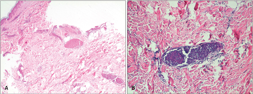
Fig. 2 (A) Skin biopsy showing deep dermis vessels with luminal occlusion due to the presence of neutrophils and fibrinoid material (H&E, ×20). (B) Detail at high magnification showing an intraluminal neutrophil thrombus in the deep dermis vessels (H&E, ×100).
Reference
-
1. Delgado-Jiménez Y, Fraga J, Fernández-Herrera J, García-Diez A. Septic vasculopathy. Actas Dermosifiliogr. 2007; 98:Suppl 1. 22–28.2. Choffray A, Flageul B, Dubertret L, Viguier M. Erysipelas-like dermatitis of the legs revealing aspergilloma of the maxillary sinus. Ann Dermatol Venereol. 2007; 134:851–854.3. Carlson JA, Chen KR. Cutaneous pseudovasculitis. Am J Dermatopathol. 2007; 29:44–55.
Article4. Legout L, Sarraz-Bournet B, D'Elia PV, Devos P, Pasquet A, Caillaux M, et al. Characteristics and prognosis in patients with prosthetic vascular graft infection: a prospective observational cohort study. Clin Microbiol Infect. 2012; 18:352–358.
Article
- Full Text Links
-
- Actions
-
Cited
- CITED
-
- Close
- Share
-
- Similar articles
-
- A Case of Cutaneous Cholesterol Embolism
- A Case of Cutaneous Cholesterol Embolism
- Mechanical Thrombectomy for Septic Embolism Secondary to Staphylococcus lugdunensis Bacteremia without Infective Endocarditis: A Case Report
- Electrocardiographic Changes in a Patient With Pulmonary Embolism and Septic Shock
- Left Atrial Myxoma Presenting with Migratory Erythematous Maculopapules
