Ann Dermatol.
2014 Aug;26(4):541-542. 10.5021/ad.2014.26.4.541.
Wells Syndrome: Response to Dapsone Therapy
- Affiliations
-
- 1Second Department of Dermatology and Department of Venereology, "A. Sygros" Hospital, Athens, Greece. kouris2007@yahoo.com
- 2Department of Pathology, "A. Sygros" Hospital, Athens, Greece.
- KMID: 2265607
- DOI: http://doi.org/10.5021/ad.2014.26.4.541
Abstract
- No abstract available.
Figure
-
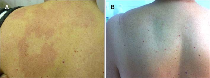
Fig. 1 (A) An indurated red-violet plaque on the upper back upon admission. (B) Clearance of eruption two weeks after the administration of dapsone 100 mg/day.

Fig. 2 (A) The presence of a "flame figure" is demonstrated in a diffuse infiltrate of eosinophils at the lower dermis (H&E, ×200). (B) Severe and diffuse dermal infiltrate of eosinophils and less in number lymphocytes and histiocytes (H&E, ×100).
Reference
-
1. Wells GC. Recurrent granulomatous dermatitis with eosinophilia. Trans St Johns Hosp Dermatol Soc. 1971; 57:46–56.2. Moossavi M, Mehregan DR. Wells' syndrome: a clinical and histopathologic review of seven cases. Int J Dermatol. 2003; 42:62–67.
Article3. Marks R. Eosinophilic cellulitis--a response to treatment with dapsone: case report. Australas J Dermatol. 1980; 21:10–12.
Article4. Moon SH, Shin MK. Bullous eosinophilic cellulitis in a child treated with dapsone. Pediatr Dermatol. 2013; 30:e46–e47.
Article5. Bozeman PM, Learn DB, Thomas EL. Inhibition of the human leukocyte enzymes myeloperoxidase and eosinophil peroxidase by dapsone. Biochem Pharmacol. 1992; 44:553–563.
Article
