A Case of Subungual Melanoma with Tumor Invasion Sparing the Nail Matrix Dermis
- Affiliations
-
- 1Department of Dermatology, Samsung Medical Center, Sungkyunkwan University School of Medicine, Seoul, Korea. dylee@skku.edu
- 2Department of Pathology, Samsung Medical Center, Sungkyunkwan University School of Medicine, Seoul, Korea.
- 3Department of Plastic Surgery, Samsung Medical Center, Sungkyunkwan University School of Medicine, Seoul, Korea.
- 4Faculty of Medicine, University of Ottawa, Ottawa, Canada.
- KMID: 2265514
- DOI: http://doi.org/10.5021/ad.2014.26.5.655
Abstract
- No abstract available.
Figure
-
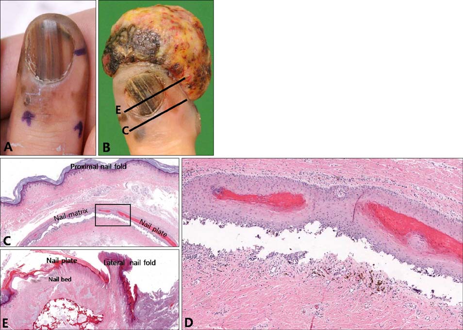
Fig. 1 (A) The patient presents with total melanonychia and dark brown macules around the 4th fingernail. (B) Six and a half months later, he shows a large tumor on the 4th finger without any definite nail deformity. Line C and line E indicate each section orientations of (C) and (E). (C) Transverse section on the proximal nail matrix shows melanoma in situ without dermal invasion. (D) High-power view of the boxed portion of (C). (E) Transverse section through the nail plate shows melanoma in situ on the nail bed and large invasive melanoma on the lateral side of the finger.
Cited by 1 articles
-
Acral Lentiginous Melanoma, Indolent Subtype Diagnosed by En Bloc Excision: A Case Report
Jungyoon Ohn, Jeong Mo Bae, Ji Soo Lim, Jong Seo Park, Hyun-Sun Yoon, Soyun Cho, Hyun-sun Park
Ann Dermatol. 2017;29(3):327-330. doi: 10.5021/ad.2017.29.3.327.
Reference
-
1. de Berker DA, André J, Baran R. Nail biology and nail science. Int J Cosmet Sci. 2007; 29:241–275.
Article2. Lee DY, Park JH, Shin HT, Yang JM, Jang KT, Kwon GY, et al. The presence and localization of onychodermis (specialized nail mesenchyme) containing onychofibroblasts in the nail unit: a morphological and immunohistochemical study. Histopathology. 2012; 61:123–130.
Article3. Tan KB, Moncrieff M, Thompson JF, McCarthy SW, Shaw HM, Quinn MJ, et al. Subungual melanoma: a study of 124 cases highlighting features of early lesions, potential pitfalls in diagnosis, and guidelines for histologic reporting. Am J Surg Pathol. 2007; 31:1902–1912.4. Izumi M, Ohara K, Hoashi T, Nakayama H, Chiu CS, Nagai T, et al. Subungual melanoma: histological examination of 50 cases from early stage to bone invasion. J Dermatol. 2008; 35:695–703.
Article5. Ruben BS. Pigmented lesions of the nail unit: clinical and histopathologic features. Semin Cutan Med Surg. 2010; 29:148–158.
Article
- Full Text Links
-
- Actions
-
Cited
- CITED
-
- Close
- Share
-
- Similar articles
-
- Three Cases of Nail Matrix Nevus in Children
- Nail sparing and sub-nail bed approach for the excision of subungual glomus tumors
- A Case of Subungual Melanoma in Situ Presenting Longitudinal Melanonychia
- Diagnosis and treatment of subungual melanoma
- A Case of Melanonychia Striata Caused by Congenital Melanocytic Nevus
