Ann Dermatol.
2014 Oct;26(5):649-650. 10.5021/ad.2014.26.5.649.
Generalized Eruptive Lentiginosis in a Healthy Elderly Man
- Affiliations
-
- 1Department of Dermatology, Chosun University Medical School, Gwangju, Korea. derm75@chosun.ac.kr
- 2Department of Neurosurgery, Chosun University Medical School, Gwangju, Korea.
- KMID: 2265510
- DOI: http://doi.org/10.5021/ad.2014.26.5.649
Abstract
- No abstract available.
Figure
-
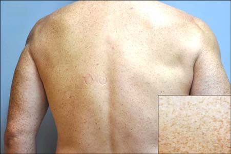
Fig. 1 Hundreds of hyperpigmented macules on the back and extremities. Inset: close-up view of lesions.
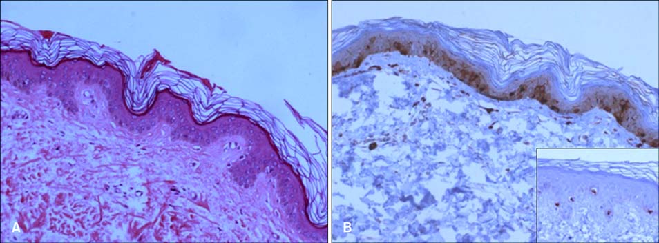
Fig. 2 (A) Pathologic features of the skin on the trunk with lentigo (H&E, ×200). (B) Proliferation of melanocytes, positive for S-100, is evident in the basal layer of the epidermis (S-100, ×200). Inset: melanocytes on Melan A staining (×200).
Reference
-
1. Uhle P, Norvell S Jr. Generalized lentiginosis. J Am Acad Dermatol. 1988; 18:444–447.
Article2. Na JI, Park KC, Youn SW. Familial eruptive lentiginosis. J Am Acad Dermatol. 2006; 55:s38–s40.
Article3. Dumas A, Petouraud C, Benoit . Lentiginose profuse et diffuse d'apparition récente chez une femmeâgée. Bull Soc Fr Dermatol Syphiligr. 1930; 40:1049–1051.4. De D, Dogra DS, Kanwar AJ, Saikia UN. Generalized eruptive lentiginosis induced by chemotherapy. Clin Exp Dermatol. 2010; 35:e113–e115.
Article5. Xing Q, Chen X, Wang M. A locus for familial generalized lentiginosis without systemic involvement maps to 3. Hum Genet. 2005; 117:154–159.
Article
- Full Text Links
-
- Actions
-
Cited
- CITED
-
- Close
- Share
-
- Similar articles
-
- Generalized Eruptive Histiocytoma
- A Case of Bilateral Segmental Neurofibromatosis Associated with Partial Unilateral Lentiginosis and Nevus of Ota
- A Case of Generalized Eruptive Syringoma
- A Case of Eruptive Vellus Hair Cyst Combined With Steatocystoma Multiplex
- A Case of Generalized Eruptive Histiocytoma in Childhood
