Ann Dermatol.
2014 Oct;26(5):639-640. 10.5021/ad.2014.26.5.639.
A Suspected Case and Literature Review of McCune-Albright Syndrome
- Affiliations
-
- 1Department of Dermatology, Eulji University Hospital, Daejeon, Korea.
- 2Department of Dermatology, The Catholic University of Korea, Seoul St. Mary's Hospital, Seoul, Korea. tykimder@catholic.ac.kr
- KMID: 2265505
- DOI: http://doi.org/10.5021/ad.2014.26.5.639
Abstract
- No abstract available.
MeSH Terms
Figure
-

Fig. 1 Solitary, brown patch with irregular border, throughout the patient's right cheek and upper eyelid with facial asymmetry. (B) Coronal computed tomography scan demonstrates heterogenous ground glass appearance at the right anterior skull, consistent with polyostotic fibrous dysplasia of the skull, right maxilla and mandible and narrowing of superior and inferior orbital canal, and compressing the right optic nerve.
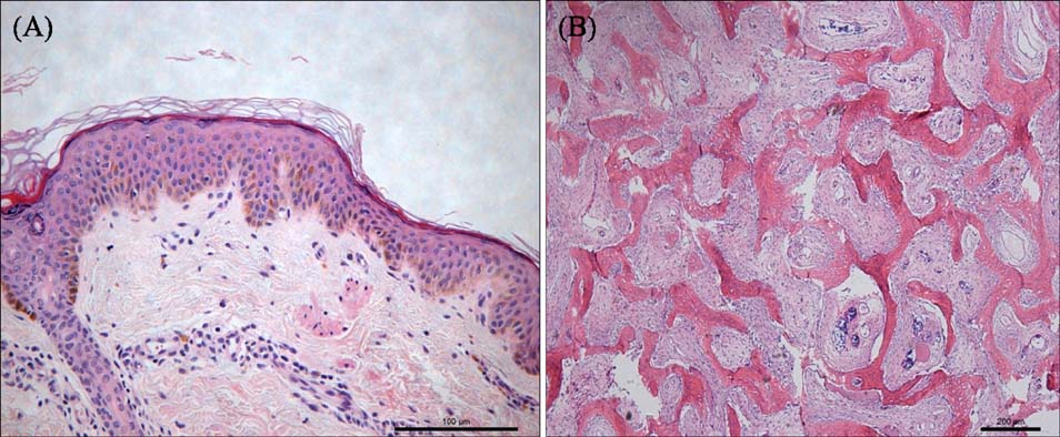
Fig. 2 (A) Hyperkeratosis, epidermal acanthosis, basal layer hyperpigmentation, and perivascular inflammatory infiltrates in the dermis (H&E, ×100). (B) Curvilinear trabeculae of woven bone admixed with fibrous tissue without nuclear atypia and mitoses (H&E, ×40).
Reference
-
1. Dumitrescu CE, Collins MT. McCune-Albright syndrome. Orphanet J Rare Dis. 2008; 3:12.
Article2. Collins MT, Singer FR, Eugster E. McCune-Albright syndrome and the extraskeletal manifestations of fibrous dysplasia. Orphanet J Rare Dis. 2012; 7:Suppl 1. S4.
Article3. Chapurlat RD, Orcel P. Fibrous dysplasia of bone and McCune-Albright syndrome. Best Pract Res Clin Rheumatol. 2008; 22:55–69.
Article4. Landau M, Krafchik BR. The diagnostic value of café-au-lait macules. J Am Acad Dermatol. 1999; 40:877–890. quiz 891-892.
Article
- Full Text Links
-
- Actions
-
Cited
- CITED
-
- Close
- Share
-
- Similar articles
-
- A case of McCune-Albright syndrome
- A Case of Ketoconazole Treatment in McCune-Albright Syndrome
- Endoscopic Decompression for Optic Neuropathy in McCune-Albright Syndrome
- A Rare Variant of Mazabraud’s Syndrome Overlapping with McCune-Albright Syndrome with a Clinical Review: A Case Report
- The McCune-Albright's syndrome: a case report and review of the literature
