A Case of Xanthoma Disseminatum Accentuating over the Eyelids
- Affiliations
-
- 1Department of Dermatology, Kyungpook National University School of Medicine, Daegu, Korea.
- 2Department of Dermatology, Medical Research Institute, Pusan National University School of Medicine, Busan, Korea. dockbs@pusan.ac.kr
- KMID: 2265376
- DOI: http://doi.org/10.5021/ad.2010.22.3.353
Abstract
- Xanthoma disseminatum (XD) is a rare, benign non-familial mucocutaneous disorder, which is a subset of non-Langerhans cell histiocytosis. It is characterized by mucocutaneous xanthomas in a disseminated form typically involving the eyelids, trunk, face, and proximal extremities and occurs in flexures and folds such as axillae and the groin. Mucosal involvement of the respiratory or gastrointestinal tracts may lead to hoarseness or intestinal obstruction from a mechanical mass effect. This paper outlines the case of a 47-year-old female with progressive yellow-to-brown confluent nodules and plaques of various sizes on her scalp, face, oral mucosa, neck, shoulder, axillary folds, and perianal area. Xanthomas accentuating over the eyelids and eyelashes led to partial obstruction of her visual field and interfered with blinking. Further, she suffered from xerophthalmia. The presentation of histopathological features including foamy histiocytes, inflammatory cells, and Touton giant cells in conjunction with her clinical findings indicated a diagnosis of XD. Evaluations for extracutaneous involvement including the central nervous system, respiratory tract, gastrointestinal tract, and bone resulted in nonspecific findings. Although she has been treated with surgical excisions, CO2 laser therapy, and oral prednisolone, new lesions are still emerging.
MeSH Terms
-
Axilla
Blinking
Central Nervous System
Extremities
Eyelashes
Eyelids
Female
Gastrointestinal Tract
Giant Cells
Groin
Histiocytes
Histiocytosis
Histiocytosis, Non-Langerhans-Cell
Hoarseness
Humans
Intestinal Obstruction
Lasers, Gas
Middle Aged
Mouth Mucosa
Neck
Prednisolone
Respiratory System
Scalp
Shoulder
Visual Fields
Xanthomatosis
Xerophthalmia
Prednisolone
Figure
-
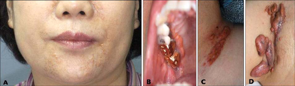
Fig. 1 Cutaneous features characterized by multiple confluent yellow to red-brown hard papules and nodules on the face (A), oral mucosa (B), neck (C), axillae (D).

Fig. 2 Xanthoma disseminatum progressively accentuating around ocular area (A) and it hindered in closing her eyes (B).

Fig. 3 Histopathologic features characterized by a mixture of histiocytes, inflammatory cells, foam cells (A: H&E, ×200), form cells and Touton giant cells (B: H&E, ×200). Immunohistochemical stainings positive for CD68 (C) and negative for S-100 (D), and CD1a (E) (×200).
Cited by 1 articles
-
Successful Treatment of Xanthoma Disseminatum with Combined Lipid Lowering Agents
Won-Jeong Kim, Hyun-Chang Ko, Byung-Soo Kim, Moon-Bum Kim
Ann Dermatol. 2012;24(3):380-382. doi: 10.5021/ad.2012.24.3.380.
Reference
-
1. Carlo G, Ruggero C. Wolff K, Goldsmith LA, Katz SI, Gilchrest BA, Paller AS, Leffell DJ, editors. Non-Langerhans cell histiocytosis. Fitzpatrick's dermatology in general medicine. 2008. 7th ed. New York: McGraw-Hill;1424–1434.2. Ozçelik U, Doğru D, Akçören Z, Coşkun T, Kaya S, Göçmen A. Xanthoma disseminatum: a child with respiratory system involvement and bronchiectasis. Pediatr Pulmonol. 2005. 39:84–87.
Article3. Kang TW, Kim SC. A case of xanthoma disseminatum presenting as pedunculating nodules and plaques. Korean J Dermatol. 2007. 45:290–293.4. Wayman LL, Margo CE. Xanthoma disseminatum with bilateral epibulbar involvement. Am J Ophthalmol. 2005. 139:557–559.
Article5. Woollons A, Darley CR. Xanthoma disseminatum: a case with hepatic involvement, diabetes insipidus and type IIb hyperlipidaemia. Clin Exp Dermatol. 1998. 23:277–280.
Article6. Varotti C, Bettoli V, Berti E, Cavicchini S, Caputo R. Xanthoma disseminatum: a case with extensive mucous membrane involvement. J Am Acad Dermatol. 1991. 25:433–436.
Article7. Calverly DC, Wismer J, Rosenthal D, deSa D, Barr RD. Xanthoma disseminatum in an infant with skeletal and marrow involvement. J Pediatr Hematol Oncol. 1995. 17:61–65.
Article8. Zak IT, Altinok D, Neilsen SS, Kish KK. Xanthoma disseminatum of the central nervous system and cranium. Am J Neuroradiol. 2006. 27:919–921.9. Büyükavci M, Selimoglu A, Yildirim U, Ertekin V, Atasoy M. Xanthoma disseminatum with hepatic involvement in a child. Pediatr Dermatol. 2005. 22:550–553.
Article10. Weiss N, Keller C. Xanthoma disseminatum: a rare normolipemic xanthomatosis. Clin Investig. 1993. 71:233–238.
Article11. Walter HB, Bernhard Z. Elder DE, Elenitsas R, Johnson BL, Murphy GF, Xu X, editors. The histiocytoses. Lever's histopathology of the skin. 2009. 10th ed. Philadelphia: Lippincott Williams & Wilkins;667–688.12. Zelger B, Cerio R, Orchard G, Fritsch P, Wilson-Jones E. Histologic and immunohistochemical study comparing xanthoma disseminatum and histiocytosis X. Arch Dermatol. 1992. 128:1207–1212.
Article13. Yoon JS, Kim YH, Lee JH, Song KY, Park JK. A case of xanthoma disseminatum. Korean J Dermatol. 1993. 31:812–816.14. Alexander AS, Turner R, Uniate L, Pearcy RG. Xanthoma disseminatum: a case report and literature review. Br J Radiol. 2005. 78:153–157.
Article15. Choi KW, Lee CY, Lee YK, Kim HS, Lee CW, Kim KH, et al. A case of xanthoma disseminatum with diabetes insipidus. Korean J Dermatol. 2008. 46:826–830.16. Seaton ED, Pillai GJ, Chu AC. Treatment of xanthoma disseminatum with cyclophosphamide. Br J Dermatol. 2004. 150:346–349.
Article17. Savaşan S, Smith L, Scheer C, Dansey R, Abella E. Successful bone marrow transplantation for life threatening xanthogranuloma disseminatum in neurofibromatosis type-1. Pediatr Transplant. 2005. 9:534–536.
Article18. Eisendle K, Linder D, Ratzinger G, Zelger B, Philipp W, Piza H, et al. Inflammation and lipid accumulation in xanthoma disseminatum: therapeutic considerations. J Am Acad Dermatol. 2008. 58:S47–S49.
Article19. Yusuf SM, Mijinyawa MS, Musa BM, Mohammed AZ. Xanthoma disseminatum in a black African woman. Int J Dermatol. 2008. 47:1145–1147.
Article
