Ann Dermatol.
2014 Dec;26(6):783-785. 10.5021/ad.2014.26.6.783.
A Case of Reticulate Acropigmentation of Kitamura Treated with 532-nm Q-Switched Nd:YAG Laser: 10 Years of Follow-Up Observation
- Affiliations
-
- 1Department of Dermatology, Samsung Medical Center, Sungkyunkwan University School of Medicine, Seoul, Korea. jooheung.lee@samsung.com
- KMID: 2264888
- DOI: http://doi.org/10.5021/ad.2014.26.6.783
Abstract
- No abstract available.
MeSH Terms
Figure
-
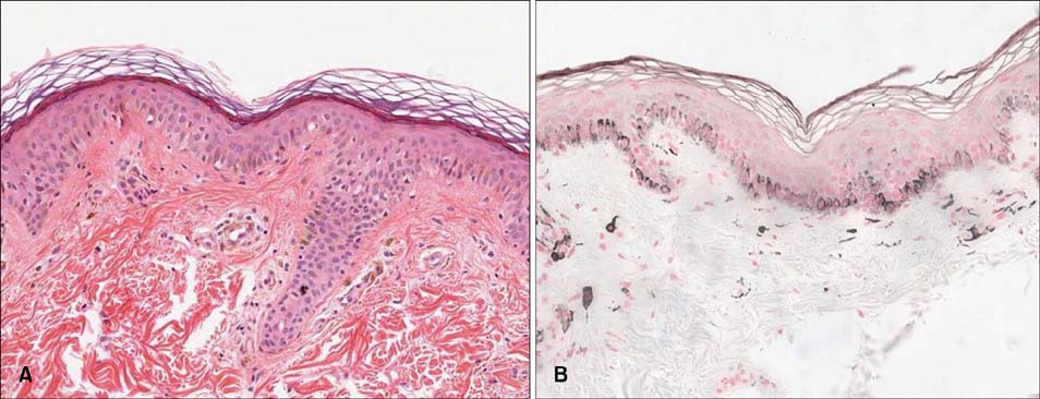
Fig. 1 (A) Lentiginous melanocytic hyperplasia and dermal melanophages with some degree of epidermal atrophy (H&E, ×100). (B) Lentiginous hyperplasia of the basal melanocytes (Fontana Masson, ×100).
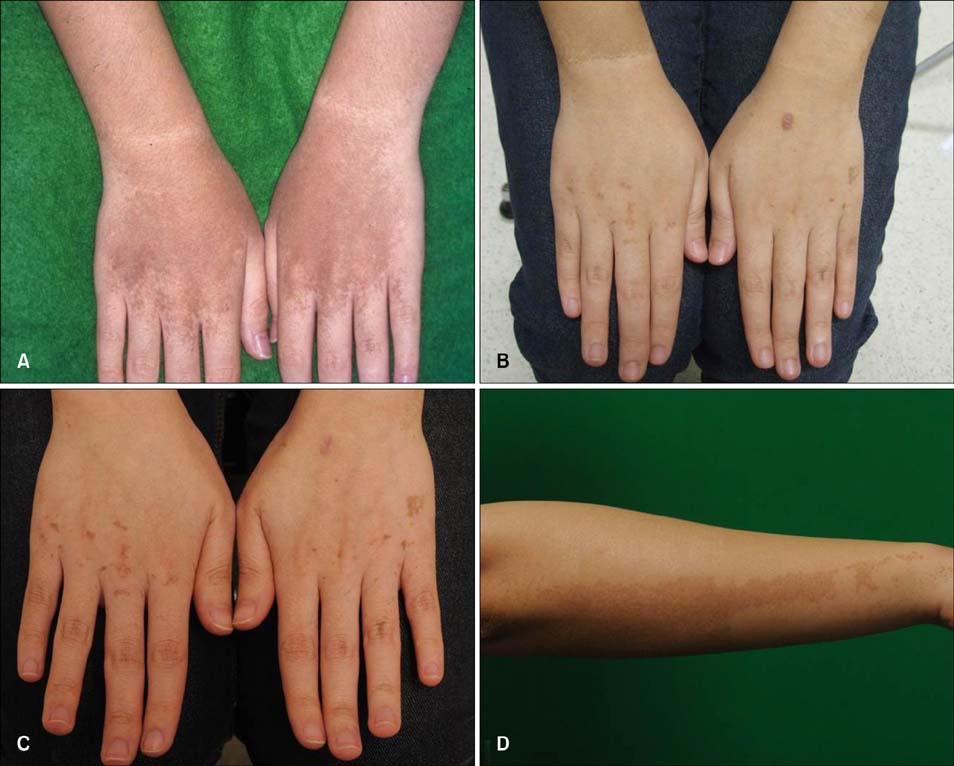
Fig. 2 (A) Reticulated brownish patches on the dorsum of both hands before treatment. (B) Significant improvement of hyperpigmentation after seven sessions of treatment. (C) Clinical improvement was maintained after 4 years without treatment. (D) Significant clinical improvement with a clear margin distinguishing the treated from the untreated area.
Reference
-
1. Kitamura K, Akamatsu S, Hirokawa K. A special form of acropigmentation: acropigmentation reticularis. Hautarzt. 1953; 4:152–156.2. Griffiths WA. Reticulate acropigmentation of Kitamura. Br J Dermatol. 1976; 95:437–443.
Article3. Gatti S, Nini G. Treatment of reticulate acropigmentation of Kitamura with azelaic acid. J Am Acad Dermatol. 1993; 29:666–667.
Article4. Fahad AS, Al Shahwan H, Bin Dayel S. Treatment of reticulated acropigmentation of Kitamura with Q-switched alexandrite laser. Int J Dermatol. 2011; 50:1150–1152.
Article
- Full Text Links
-
- Actions
-
Cited
- CITED
-
- Close
- Share
-
- Similar articles
-
- Acropigmentation Symmetrica of Dohi Treated with the Q-switched Alexandrite Laser
- A Case of Reticulate Acropigmentation of Kitamura
- A Case of Reticulate Acropigmentation of Kitamura with Hyperpigmented Macules on the Flexural Areas
- 532-nm Q-switched Nd:YAG laser treatment for linear porokeratosis in Republic of Korea: a case report
- Two Cases of Reticulate Acropigmentation of Kitamura
