Recurrent Acral Lentiginous Melanoma In Situ Suggesting the Field Cell Theory
- Affiliations
-
- 1Department of Dermatology, Seoul National University College of Medicine, Seoul, Korea. khcho@snu.ac.kr
- 2Institute of Human-Environment Interface Biology, Seoul National University, Seoul, Korea.
- 3Laboratory of Cutaneous Aging and Hair Research, Biomedical Research Institute, Seoul National University Hospital, Seoul, Korea.
- KMID: 2264886
- DOI: http://doi.org/10.5021/ad.2014.26.6.779
Abstract
- No abstract available.
MeSH Terms
Figure
-
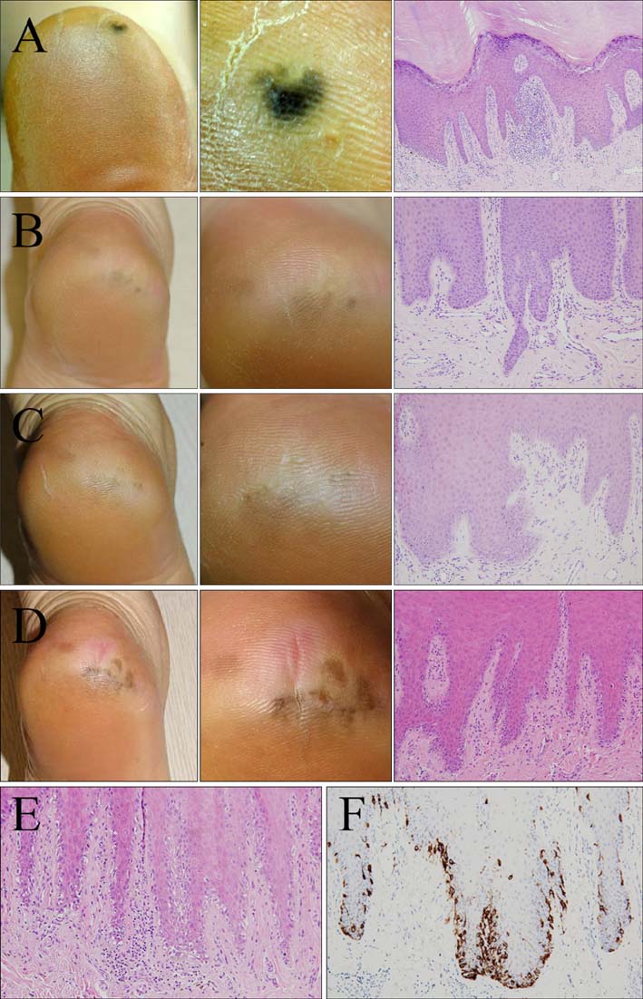
Fig. 1 Acral lentiginous melanoma in situ. Clinical and histopathological feature. (A) Initial presentation 8 years ago. (B) Second presentation 6 years ago. (C) Third presentation 4 years ago. (D) Last presentation. Left, gross picture of the lesion; center, close-up picture of the lesion showing multiple dark brown to black pigmented enlarged patches with irregular border and variegated color on the left heel; right, histopathologic feature demonstrating bland proliferation of scattered melanocytes without marked atypia or dermal invasion (A~D: H&E, ×200). (E) Lentiginous hyperplasia of melanocytes was prominent only in the epidermis (H&E, ×200). (F) Melan-A staining showed atypical melanocytes confined to the epidermis (immunoperoxidase, ×200).
Reference
-
1. Arrington JH 3rd, Reed RJ, Ichinose H, Krementz ET. Plantar lentiginous melanoma: a distinctive variant of human cutaneous malignant melanoma. Am J Surg Pathol. 1977; 1:131–143.2. Nogita T, Wong TY, Ohara K, Mizushima J, Mihm MC Jr, Kawashima M. Atypical melanosis of the foot. A report of three cases in Japanese populations. Arch Dermatol. 1994; 130:1042–1045.
Article3. Kilinc Karaarslan I, Akalin T, Unal I, Ozdemir F. Atypical melanosis of the foot showing a dermoscopic feature of the parallel ridge pattern. J Dermatol. 2007; 34:56–59.
Article4. Chiu HH, Hu SC, Ke CL, Cheng ST. Dermoscopy identifies histopathologically indiscernible malignant lesion of atypical melanosis of the foot, an early lesion of acral lentiginous melanoma in situ. Dermatol Surg. 2008; 34:979–983.
Article5. Takata M, Murata H, Saida T. Molecular pathogenesis of malignant melanoma: a different perspective from the studies of melanocytic nevus and acral melanoma. Pigment Cell Melanoma Res. 2010; 23:64–71.
Article
- Full Text Links
-
- Actions
-
Cited
- CITED
-
- Close
- Share
-
- Similar articles
-
- Acral Lentiginous Melanoma in situ
- Acral Lentiginous Melanoma Developing during Long-standing Atypical Melanosis: Usefulness of Dermoscopy for Detection of Early Acral Melanoma
- Progression from Acral Lentiginous Melanoma in situ to Invasive Acral Lentiginous Melanoma
- A Case of Acral Lentiginous Melanoma
- Acral Lentiginous Melanoma, Indolent Subtype Diagnosed by En Bloc Excision: A Case Report
