Ann Dermatol.
2015 Feb;27(1):109-110. 10.5021/ad.2015.27.1.109.
Interferon-Gamma Release Assay in a Patient with Tuberculosis Verrucosa Cutis
- Affiliations
-
- 1Department of Dermatology, VHS Medical Center, Seoul, Korea. jade1954@hanmail.net
- 2Department of Dermatology, Wonkwang University, Iksan, Korea.
- KMID: 2264854
- DOI: http://doi.org/10.5021/ad.2015.27.1.109
Abstract
- No abstract available.
Figure
-

Fig. 1 A 7×8 cm annular erythematous plaque with central clearing and hyperpigmentation in the left inner thigh.
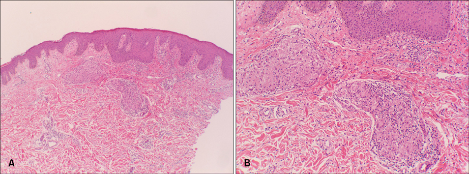
Fig. 2 (A) Parakeratosis, acanthosis in epidermis and naked granulomas in the upper to mid-dermis (H&E, ×40). (B) Naked granulomas composed of epithelioid cells (H&E, ×200).
Cited by 1 articles
-
Multiple Skin Colored Nodules on both Legs in Patient with Positive QuantiFERON®-TB Gold Test
Mi Soo Choi, Seung Phil Hong, Byung Cheol Park, Myung Hwa Kim
Ann Dermatol. 2017;29(1):95-99. doi: 10.5021/ad.2017.29.1.95.
Reference
-
1. Damevska K, Gocev G. Multifocal tuberculosis verrucosa cutis of 60 years duration. Int J Infect Dis. 2013; 17:e1266–e1267.
Article2. Sethi A. Fitzpatrick's dermatology in general medicine. In : Goldsmith LA, Katz SI, Gilchrest BA, Paller AS, Leffell DJ, Wolff K, editors. Tuberculosis and infections with atypical mycobacteria. 8th ed. New York: McGraw-Hill;2012. p. 2225–2241.3. Kardos M, Kimball AB. Time for a change? Updated guidelines using interferon gamma release assays for detection of latent tuberculosis infection in the office setting. J Am Acad Dermatol. 2012; 66:148–152.
Article4. Koh HY, Tay LK, Pang SM, Ong BH. Changing the way we diagnose tuberculids with interferon gamma release assays. Australas J Dermatol. 2012; 53:73–75.
Article
- Full Text Links
-
- Actions
-
Cited
- CITED
-
- Close
- Share
-
- Similar articles
-
- Tuberculosis Verrucosa Cutis in a Patient with Pulmonary Tuberculosis
- A Case of Giant Tuberculosis Verrucosa Cutis Associated with Bladder Cancer
- A Case of Tuberculosis Verrucosa Cutis with Ulcer in a Patient with Pulmonary Tuberculosis
- Tuberculosis Verrucosa Cutis Confirmed by Culture
- A Case of Tuberculosis Verrucosa Cutis
