Ann Dermatol.
2015 Apr;27(2):226-227. 10.5021/ad.2015.27.2.226.
Preoperative Evaluation of a Subungual Glomus Tumor Case Using Multidetector Computed Tomography Angiography
- Affiliations
-
- 1Department of Radiology, Affiliated Hospital of Guangdong Medical College, Zhanjiang, China.
- 2Department of Dermatology, Affiliated Hospital of Guangdong Medical College, Zhanjiang, China. ymfan1963@163.com
- 3Department of Ultrasound, Affiliated Hospital of Guangdong Medical College, Zhanjiang, China.
- KMID: 2264822
- DOI: http://doi.org/10.5021/ad.2015.27.2.226
Abstract
- No abstract available.
Figure
-
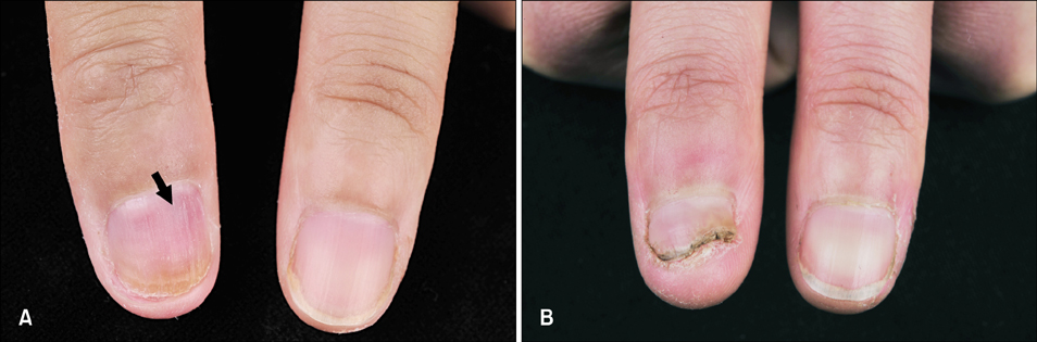
Fig. 1 Subungual glomus tumor on the distal part of the right thumb. (A) A 6×4-mm purplish flush (arrow) with distal yellowish discoloration on the right thumb. (B) No nail abnormality present at 4 months postoperation.
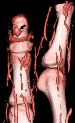
Fig. 2 Volume-rendered image of the vascular reconstruction displaying a hypervascularized nodule (arrow) overlying the distal phalanx of the right thumb.
Reference
-
1. Netscher DT, Aburto J, Koepplinger M. Subungual glomus tumor. J Hand Surg Am. 2012; 37:821–823.
Article2. Hamdi MF. Glomus tumour of fingertip: report of eight cases and literature review. Musculoskelet Surg. 2011; 95:237–240.
Article3. Matsunaga A, Ochiai T, Abe I, Kawamura A, Muto R, Tomita Y, et al. Subungual glomus tumour: evaluation of ultrasound imaging in preoperative assessment. Eur J Dermatol. 2007; 17:67–69.4. Theumann NH, Goettmann S, Le Viet D, Resnick D, Chung CB, Bittoun J, et al. Recurrent glomus tumors of fingertips: MR imaging evaluation. Radiology. 2002; 223:143–151.
Article5. Li K, Moon SE. Letter: creating preoperative images of subungual glomus tumors. Dermatol Surg. 2008; 34:1296–1297.
