Primary cutaneous B-cell lymphoblastic lymphoma in an elderly man
- Affiliations
-
- 1Department of Internal Medicine, Korea Cancer Center Hospital, Korea Institute of Radiological and Medical Sciences, Seoul, Korea. hyejin@kcch.re.kr
- 2Department of Pathology, Korea Cancer Center Hospital, Korea Institute of Radiological and Medical Sciences, Seoul, Korea.
- KMID: 2251991
- DOI: http://doi.org/10.5045/kjh.2011.46.4.283
Abstract
- Precursor B-cell lymphoblastic lymphoma (B-LBL) is an uncommon high-grade neoplasm of immature B cells. It occurs predominantly in childhood with extranodal involvement such as skin and bone. Therefore, primary cutaneous involvement in elderly adults is a very rare manifestation of B-LBL. Here, we report a 78-year-old man with B-LBL presenting as a single cutaneous lesion which was immunohistochemically positive for leukocyte common antigen (LCA), CD79a, paired box 5 (PAX5), B cell lymphoma-2 (bcl-2), and terminal deoxynucleotidyl transferase (TdT) staining, but was without systemic involvement. The patient was treated using cyclophosphamide, adriamycin, vincristine, and prednisolone (CHOP), and achieved complete response (CR) at the first response assessment conducted after 3 CHOP cycles. After an additional cycle of CHOP treatment, radiotherapy was administered at a total dose of 3,600 cGy over 4 weeks. At the 21-month follow-up, he had maintained CR.
MeSH Terms
-
Adult
Aged
Antigens, CD45
B-Lymphocytes
Cyclophosphamide
DNA Nucleotidylexotransferase
Doxorubicin
Follow-Up Studies
Humans
Precursor B-Cell Lymphoblastic Leukemia-Lymphoma
Precursor Cell Lymphoblastic Leukemia-Lymphoma
Precursor Cells, B-Lymphoid
Prednisolone
Skin
Vincristine
Antigens, CD45
Cyclophosphamide
DNA Nucleotidylexotransferase
Doxorubicin
Prednisolone
Vincristine
Figure
-
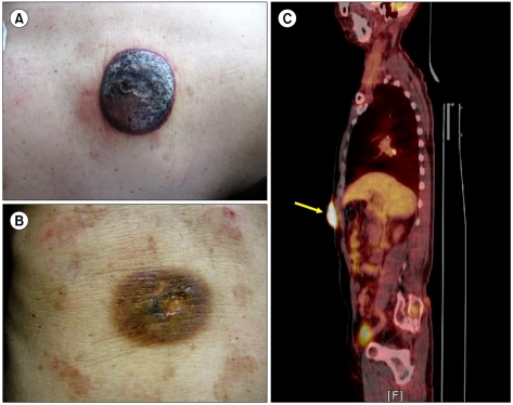
Fig. 1 (A) At diagnosis, a 50×60 mm non-tender, non-itching, and dark red tumor with smooth surface was present on the right upper abdominal quadrant. (B) After the 3rd session of cyclophosphamide, adriamycin, vincristine, and prednisolone (CHOP) chemotherapy, the tumor disappeared by gross examination. (C) 18F-fluorodeoxyglucose (FDG) positron emission tomography (PET)/computed tomography (CT) scan shows a single hypermetabolic lesion (arrow) in the abdominal wall (standardized uptake value [SUV]=12.8).
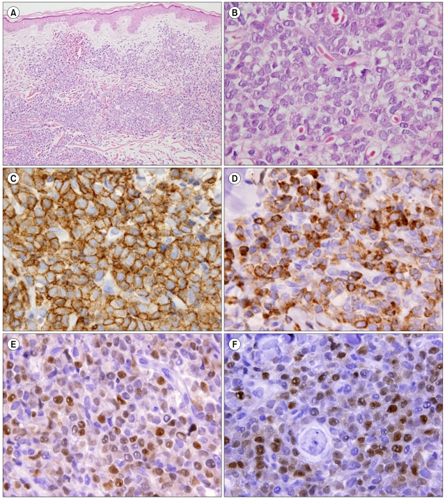
Fig. 2 (A) Microscopically, skin biopsy specimen revealed diffuse infiltration of small to intermediate lymphoid cells in the dermis and subcutaneous fat (hematoxylin & eosin [H&E], original magnification ×200). (B) High magnification shows monotonous tumor cells with slightly convoluted nuclei with fine chromatin and occasionally nucleolus and scant cytoplasm (H&E, original magnification ×1,000). (C-F) Photographs of immunohistochemical staining. Most tumor cells are positive for leukocyte common antigen (LCA) (C), CD79a (D), paired box 5 (PAX5) (E), and terminal deoxynucleotidyl transferase (TdT) (F).
Reference
-
1. The Non-Hodgkin's Lymphoma Classification Project. A clinical evaluation of the International Lymphoma Study Group classification of non-Hodgkin's lymphoma. The Non-Hodgkin's Lymphoma Classification Project. Blood. 1997; 89:3909–3918. PMID: 9166827.2. Pui CH, Behm FG, Crist WM. Clinical and biologic relevance of immunologic marker studies in childhood acute lymphoblastic leukemia. Blood. 1993; 82:343–362. PMID: 8329694.
Article3. Grogan T, Spier C, Wirt DP, et al. Immunologic complexity of lymphoblastic lymphoma. Diagn Immunol. 1986; 4:81–88. PMID: 3086016.4. Maitra A, McKenna RW, Weinberg AG, Schneider NR, Kroft SH. Precursor B-cell lymphoblastic lymphoma. A study of nine cases lacking blood and bone marrow involvement and review of the literature. Am J Clin Pathol. 2001; 115:868–875. PMID: 11392884.5. Lyu CJ, Kang IJ, Koo HH, et al. Epidemiology and clinical outcomes in children with malignant lymphoma in Korea: retrospective study. Korean J Pediatr Hematol Oncol. 2004; 11:153–163.6. Kang YK, Kim BS, Kim TW, et al. Clinicopathologic characteristics of Korean non-Hodgkin's lymphomas based on REAL classification. J Korean Cancer Assoc. 1999; 31:641–652.7. Lin P, Jones D, Dorfman DM, Medeiros LJ. Precursor B-cell lymphoblastic lymphoma: a predominantly extranodal tumor with low propensity for leukemic involvement. Am J Surg Pathol. 2000; 24:1480–1490. PMID: 11075849.8. Shafer D, Wu H, Al-Saleem T, et al. Cutaneous precursor B-cell lymphoblastic lymphoma in 2 adult patients: clinicopathologic and molecular cytogenetic studies with a review of the literature. Arch Dermatol. 2008; 144:1155–1162. PMID: 18794461.
Article9. Muljono A, Graf NS, Arbuckle S. Primary cutaneous lymphoblastic lymphoma in children: series of eight cases with review of the literature. Pathology. 2009; 41:223–228. PMID: 19291533.
Article10. Kawakami T, Kimura S, Kinoshita A, Kondo K, Soma Y. Precursor B-cell lymphoblastic lymphoma with only cutaneous involvement. Acta Derm Venereol. 2009; 89:540–541. PMID: 19734992.
Article11. Swerdlow SH, Campo E, Harris NL, editors. WHO classification of tumours of haematopoietic and lymphoid tissues. 2008. 4th ed. Lyon, France: IARC Press;p. 168–170.12. Hashimoto M, Yamashita Y, Mori N. Immunohistochemical detection of CD79a expression in precursor T cell lymphoblastic lymphoma/leukaemias. J Pathol. 2002; 197:341–347. PMID: 12115880.
Article13. Soslow RA, Baergen RN, Warnke RA. B-lineage lymphoblastic lymphoma is a clinicopathologic entity distinct from other histologically similar aggressive lymphomas with blastic morphology. Cancer. 1999; 85:2648–2654. PMID: 10375114.
Article
