Radiation sigmoiditis mimicking sigmoid colon cancer after radiation therapy for cervical cancer: the implications of three-dimensional image-based brachytherapy planning
- Affiliations
-
- 1Department of Radiation Oncology, Samsung Medical Center, Sungkyunkwan University School of Medicine, Seoul, Korea. sj5201.huh@samsung.net
- KMID: 2245179
- DOI: http://doi.org/10.3802/jgo.2012.23.3.197
Abstract
- External-beam radiation therapy with intracavitary high-dose-rate brachytherapy is the standard treatment modality for advanced cervical cancer; however, late gastrointestinal complications are a major concern after radiotherapy. While radiation proctitis is a well-known side effect and radiation oncologists make an effort to reduce it, the sigmoid colon is often neglected as an organ at risk. Herein, we report two cases of radiation sigmoiditis mimicking sigmoid colon cancer after external-beam radiation therapy with intracavitary high-dose-rate brachytherapy for uterine cervical cancer with dosimetric consideration.
Figure
-
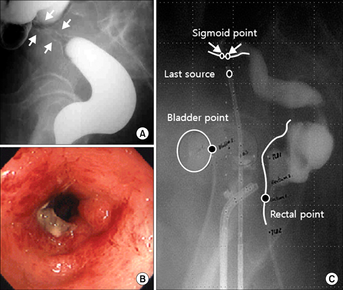
Fig. 1 Case 1: Lateral view (A) of a barium enema study shows a stricture (between arrows) of the sigmoid colon and endoscopy shows luminal narrowing and friable hyperemic nodular mucosal changes (B). A close sigmoid-to-tandem distance may cause a high sigmoid dose, which could lead to the development of sigmoiditis (C).

Fig. 2 Case 2: Endoscopy revealed luminal narrowing, hemorrhage and hyperemic mucosal nodularities with mucosal edema of the sigmoid colon (A). A CT scan of the abdomen and pelvis showed diffuse submucosal thickening of the sigmoid wall (arrow) infiltrating into the adjacent mesenteric fat, but no evidence of malignancy was seen (B).
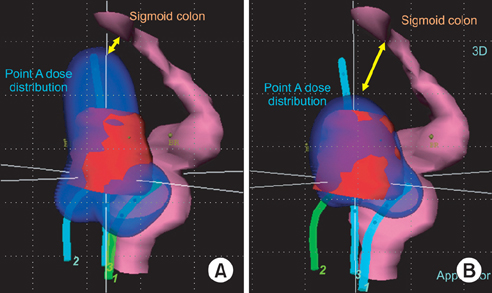
Fig. 3 Treatment planning with three dimensional (3D) image-based brachytherapy. The reconstruction image of dose distribution (blue) using traditional planning based on point A (A) shows a closer proximity to the sigmoid colon compared to 3D planning (B). applicator, sky blue; clinical target volume, red; rectum and sigmoid colon, purple.
Reference
-
1. Perez CA, Grigsby PW, Lockett MA, Chao KS, Williamson J. Radiation therapy morbidity in carcinoma of the uterine cervix: dosimetric and clinical correlation. Int J Radiat Oncol Biol Phys. 1999. 44:855–866.2. Perez CA, Breaux S, Bedwinek JM, Madoc-Jones H, Camel HM, Purdy JA, et al. Radiation therapy alone in the treatment of carcinoma of the uterine cervix. II. Analysis of complications. Cancer. 1984. 54:235–246.3. Syed AM, Puthawala AA, Abdelaziz NN, el-Naggar M, Disaia P, Berman M, et al. Long-term results of low-dose-rate interstitial-intracavitary brachytherapy in the treatment of carcinoma of the cervix. Int J Radiat Oncol Biol Phys. 2002. 54:67–78.4. Ramirez PT, Levenback C, Burke TW, Eifel P, Wolf JK, Gershenson DM. Sigmoid perforation following radiation therapy in patients with cervical cancer. Gynecol Oncol. 2001. 82:150–155.5. Galland RB, Spencer J. The natural history of clinically established radiation enteritis. Lancet. 1985. 1:1257–1258.6. Holloway CL, Racine ML, Cormack RA, O'Farrell DA, Viswanathan AN. Sigmoid dose using 3D imaging in cervical-cancer brachytherapy. Radiother Oncol. 2009. 93:307–310.7. Kim RY, Shen S, Duan J. Image-based three-dimensional treatment planning of intracavitary brachytherapy for cancer of the cervix: dose-volume histograms of the bladder, rectum, sigmoid colon, and small bowel. Brachytherapy. 2007. 6:187–194.8. Potter R, Haie-Meder C, Van Limbergen E, Barillot I, De Brabandere M, Dimopoulos J, et al. Recommendations from gynaecological (GYN) GEC ESTRO working group (II): concepts and terms in 3D image-based treatment planning in cervix cancer brachytherapy-3D dose volume parameters and aspects of 3D image-based anatomy, radiation physics, radiobiology. Radiother Oncol. 2006. 78:67–77.9. Lanciano RM, Martz K, Montana GS, Hanks GE. Influence of age, prior abdominal surgery, fraction size, and dose on complications after radiation therapy for squamous cell cancer of the uterine cervix: a patterns of care study. Cancer. 1992. 69:2124–2130.
- Full Text Links
-
- Actions
-
Cited
- CITED
-
- Close
- Share
-
- Similar articles
-
- Who Really Benefits from 3D-Based Planning of Brachytherapy for Cervical Cancer?
- Treatment Planning Software for High Dose Rate Remote Afterloading Brachytherapy of Uterine Cervical Cancer
- The using of megavoltage computed tomography in image-guided brachytherapy for cervical cancer: a case report
- Radiation therapy of cervical cancer: Current status in Korea and recent developments
- Dose comparison between prescription methods according to anatomical variations in intracavitary brachytherapy for cervical cancer
