Intrahepatic and extrahepatic intraductal papillary neoplasms of bile duct
- Affiliations
-
- 1Department of Surgery, Kosin University Gospel Hospital, Busan, Korea. yoonmhj@dreamwiz.com
- KMID: 2243171
- DOI: http://doi.org/10.14701/kjhbps.2013.17.1.48
Abstract
- There has been an increase in the number of reported cases of biliary neoplasm of the hepatobiliary system characterized by markedly dilated and multifocal papillary epithelial lesions of the bile ducts or cystic biliary lesions with or without mucin secretion, and mucinous lesions or tumors, possibly due to recent advances in radiological diagnosis. This lesion of the bile duct is believed to show a better clinical course than non-papillary biliary neoplasm. Therefore, the early recognition and treatment is important. We report two cases of intrahepatic and extrahepatic intraductal papillary neoplasm of the bile duct that were completely resected.
Keyword
MeSH Terms
Figure
-
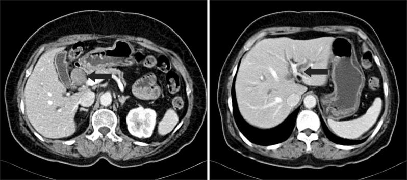
Fig. 1 Contrast-enhanced CT showing a 3.5×2.5 cm sized rounded soft tissue mass (black arrow) in the proximal common hepatic duct (CHD) and dilated bile ducts (black arrow).
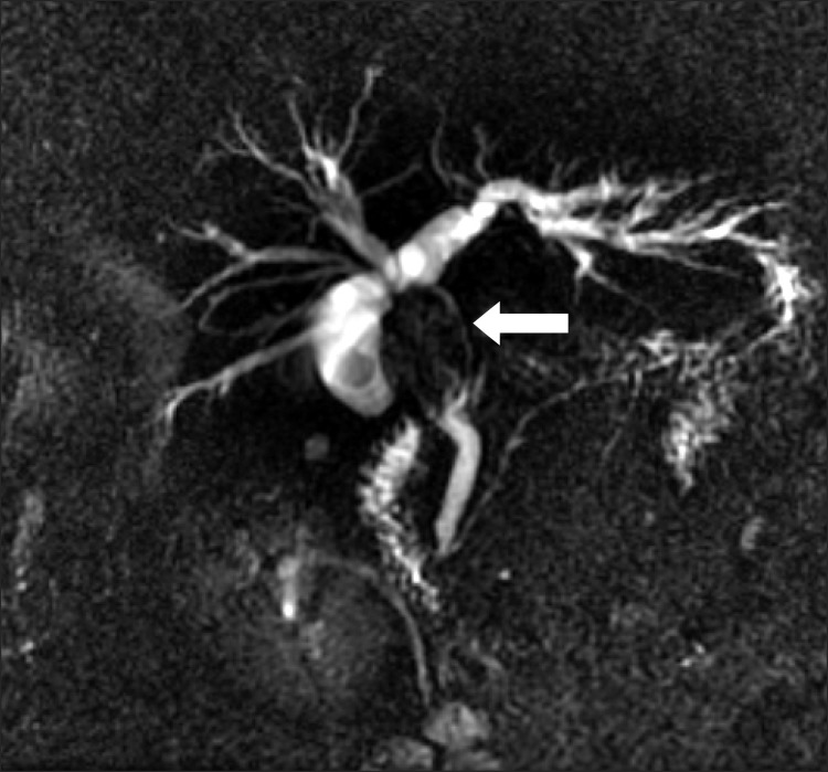
Fig. 2 MRI showing 2.6 cm by 3.9 cm sized signal void lesion in the proximal common duct and the dilatations in both IHDs (white arrow).
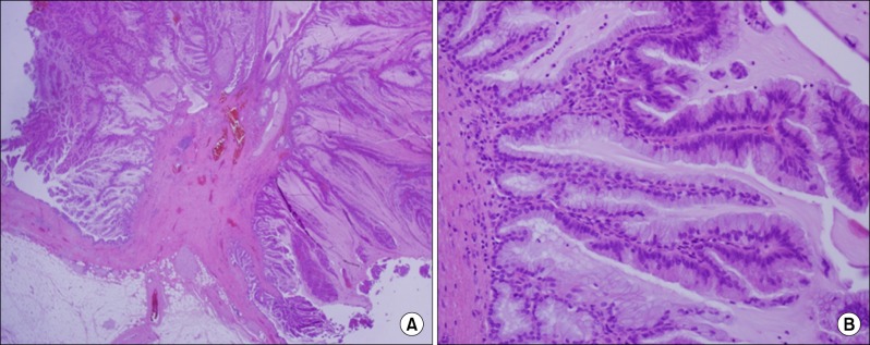
Fig. 3 A microscopic view of papillary growing neoplasm of proximal bile duct composed of elongated villotubulor structure of epithelial cells with extracellular mucin production. (A) H&E, ×40, villotubulor structure of epitherial cells; (B) H&E, ×200, multiple ductules with tall columnar mucinous epithelial cells.
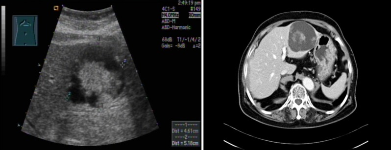
Fig. 4 Ultrasound examination showing 7.5×6 cm sized complex cystic mass in left lateral segment of liver with internal solid portion. Contrast-enhanced CT showing a well-defined 7.5×6 cm sized cystic mass with internal debris in the left lateral segment of liver.
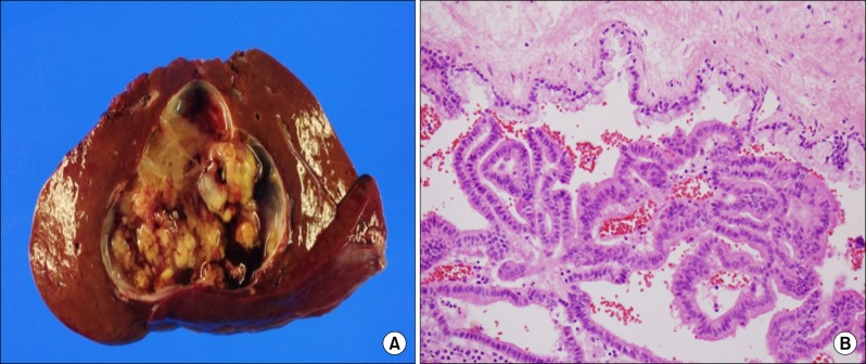
Fig. 5 A gross (A) and microscopic (B) view of papillary growing intraluminal mass. (A) An encapsulated multilobular cystic mass, filled with clear mucinous fluid. The inner surface of cyst shows multifocal papillary projections. The cut surface of the lesion is heterogeneously solid, yellowish tan and granular. (B) H&E, ×200, Papillary epithelial mass lined by relatively well-oriented columnar shaped epithelial cells with mild nuclear atypism. The cystic wall lined by single layer of mucous cells supported by loose fibrosis without ovarian stroma.
Reference
-
1. Chen TC, Nakanuma Y, Zen Y, et al. Intraductal papillary neoplasia of the liver associated with hepatolithiasis. Hepatology. 2001; 34:651–658. PMID: 11584359.
Article2. Nakanuma Y, Sasaki M, Ishikawa A, et al. Biliary papillary neoplasm of the liver. Histol Histopathol. 2002; 17:851–861. PMID: 12168796.3. Sudo Y, Harada K, Tsuneyama K, et al. Oncocytic biliary cystadenocarcinoma is a form of intraductal oncocytic papillary neoplasm of the liver. Mod Pathol. 2001; 14:1304–1309. PMID: 11743055.
Article4. Yeh TS, Tseng JH, Chiu CT, et al. Cholangiographic spectrum of intraductal papillary mucinous neoplasm of the bile ducts. Ann Surg. 2006; 244:248–253. PMID: 16858187.
Article5. Zen Y, Sasaki M, Fujii T, et al. Different expression patterns of mucin core proteins and cytokeratins during intrahepatic cholangiocarcinogenesis from biliary intraepithelial neoplasia and intraductal papillary neoplasm of the bile duct-an immunohistochemical study of 110 cases of hepatolithiasis. J Hepatol. 2006; 44:350–358. PMID: 16360234.
Article6. Albores-Saavedra J, Murakata L, Krueger JE, et al. Noninvasive and minimally invasive papillary carcinomas of the extrahepatic bile ducts. Cancer. 2000; 89:508–515. PMID: 10931449.
Article7. Nakanuma Y, Harada K, Ishikawa A, et al. Anatomic and molecular pathology of intrahepatic cholangiocarcinoma. J Hepatobiliary Pancreat Surg. 2003; 10:265–281. PMID: 14598145.
Article8. Okamoto A, Tsuruta K, Matsumoto G, et al. Papillary carcinoma of the extrahepatic bile duct: characteristic features and implications in surgical treatment. J Am Coll Surg. 2003; 196:394–401. PMID: 12648691.
Article9. Sano T, Kamiya J, Nagino M, et al. Macroscopic classification and preoperative diagnosis of intrahepatic cholangiocarcinoma in Japan. J Hepatobiliary Pancreat Surg. 1999; 6:101–107. PMID: 10398895.
Article10. Lee SS, Kim MH, Lee SK, et al. Clinicopathologic review of 58 patients with biliary papillomatosis. Cancer. 2004; 100:783–793. PMID: 14770435.
Article11. Charre L, Boillot O, Goffette P, et al. Long-term survival after isolated liver transplantation for intrahepatic biliary papillomatosis. Transpl Int. 2006; 19:249–252. PMID: 16441776.
Article12. Jang GW, Hwang S, Lee YJ, et al. Clinicopathological features of the intraductal papillary neoplasms of the intrahepatic bile duct. Korean J Hepatobiliary Pancreat Surg. 2012; 16:138–141.
Article13. Zen Y, Fujii T, Itatsu K, et al. Biliary papillary tumors share pathological features with intraductal papillary mucinous neoplasm of the pancreas. Hepatology. 2006; 44:1333–1343. PMID: 17058219.
Article14. Lim JH, Kim YI, Park CK. Intraductal mucosal-spreading mucin-producing peripheral cholangiocarcinoma of the liver. Abdom Imaging. 2000; 25:89–92. PMID: 10652930.
Article15. Yoon KH, Ha HK, Kim CG, et al. Malignant papillary neoplasms of the intrahepatic bile ducts: CT and histopathologic features. AJR Am J Roentgenol. 2000; 175:1135–1139. PMID: 11000178.16. Kim TK, Choi BI, Han JK, et al. Peripheral cholangiocarcinoma of the liver: two-phase spiral CT findings. Radiology. 1997; 204:539–543. PMID: 9240550.
Article17. Kokubo T, Itai Y, Ohtomo K, et al. Mucin-hypersecreting intrahepatic biliary neoplasms. Radiology. 1988; 168:609–614. PMID: 2841715.
Article18. Kawakatsu M, Vilgrain V, Zins M, et al. Radiologic features of papillary adenoma and papillomatosis of the biliary tract. Abdom Imaging. 1997; 22:87–90. PMID: 9000364.
Article19. Yeh CN, Jan YY, Yeh TS, et al. Hepatic resection of the intraductal papillary type of peripheral cholangiocarcinoma. Ann Surg Oncol. 2004; 11:606–611. PMID: 15172934.
Article20. Yeh TS, Tseng JH, Chen TC, et al. Characterization of intrahepatic cholangiocarcinoma of the intraductal growth-type and its precursor lesions. Hepatology. 2005; 42:657–664. PMID: 16116640.
Article21. Qin XL, Wang ZR, Shi JS, et al. Utility of serum CA19-9 in diagnosis of cholangiocarcinoma: in comparison with CEA. World J Gastroenterol. 2004; 10:427–432. PMID: 14760772.
Article22. Devaney K, Goodman ZD, Ishak KG. Hepatobiliary cystadenoma and cystadenocarcinoma. A light microscopic and immunohistochemical study of 70 patients. Am J Surg Pathol. 1994; 18:1078–1091. PMID: 7943529.23. Martin RC, Klimstra DS, Schwartz L, et al. Hepatic intraductal oncocytic papillary carcinoma. Cancer. 2002; 95:2180–2187. PMID: 12412172.
Article
- Full Text Links
-
- Actions
-
Cited
- CITED
-
- Close
- Share
-
- Similar articles
-
- Extrahepatic Bile Duct Duplication with Intraductal Papillary Neoplasm: A Case Report
- Characterization of Intraductal Papillary Neoplasm of the Bile Duct with Respect to the Histopathologic Similarities to Pancreatic Intraductal Papillary Mucinous Neoplasm
- A Case of Multiple Papillary Adenocarcinoma of the Extrahepatic Bile Duct : Findings of ERCP
- Photodynamic Therapy Followed by Left Hepatectomy Used to Treat an Intraductal Papillary Mucinous Neoplasm of the Bile Duct
- Radiological Spectrum of Intraductal Papillary Tumors of the Bile Ducts
