Focal Fatty Sparing of the Liver
- Affiliations
-
- 1Department of Internal Medicine, Hanyang University College of Medicine, Seoul, Korea. noshin@hanyang.ac.kr
- KMID: 2234042
- DOI: http://doi.org/10.4166/kjg.2014.63.6.382
Abstract
- No abstract available.
MeSH Terms
Figure
-
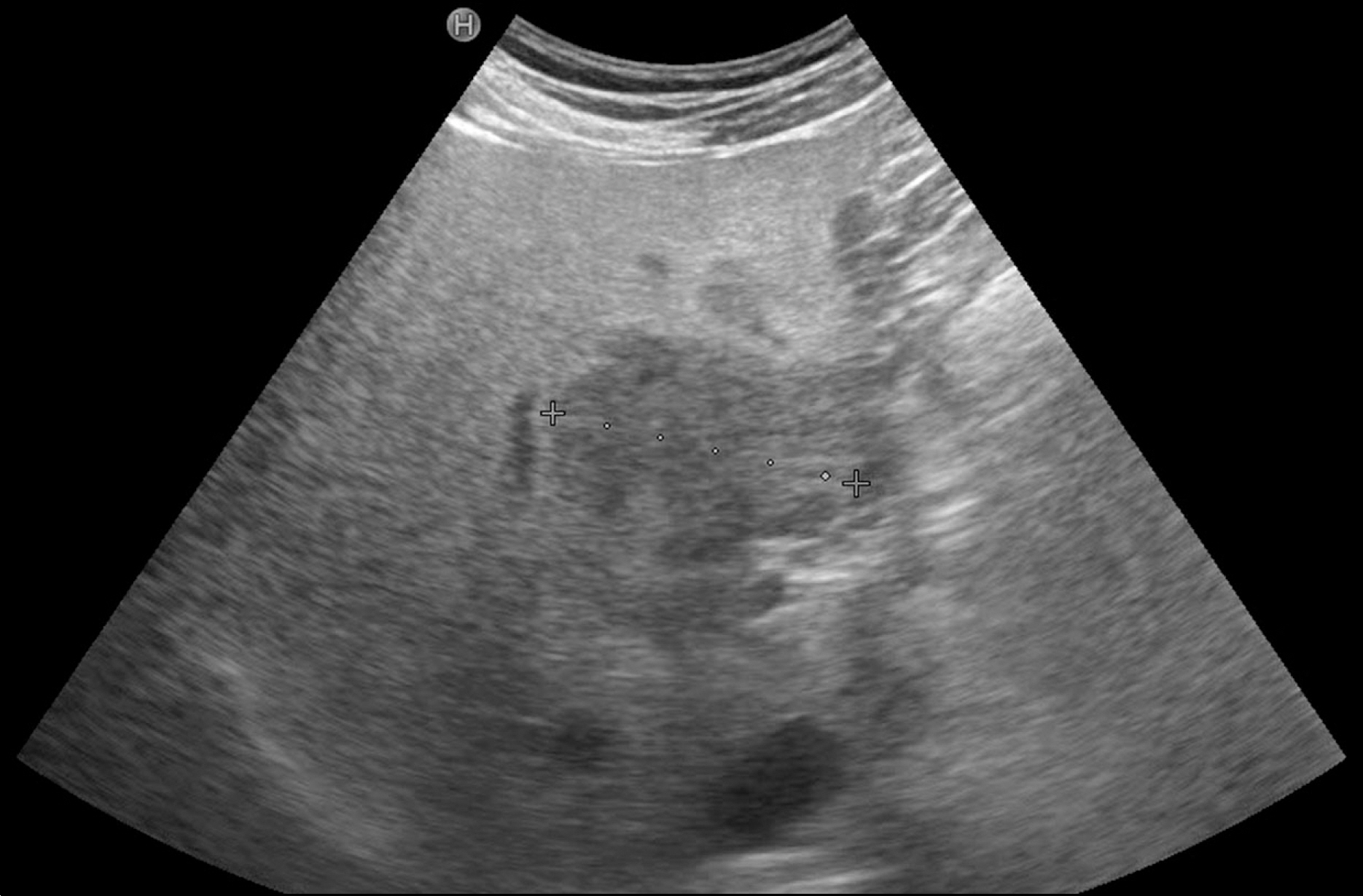
Fig. 1. Abdominal ultrasonography shows hypoechoic mass with irregular margin at segment IV of liver.
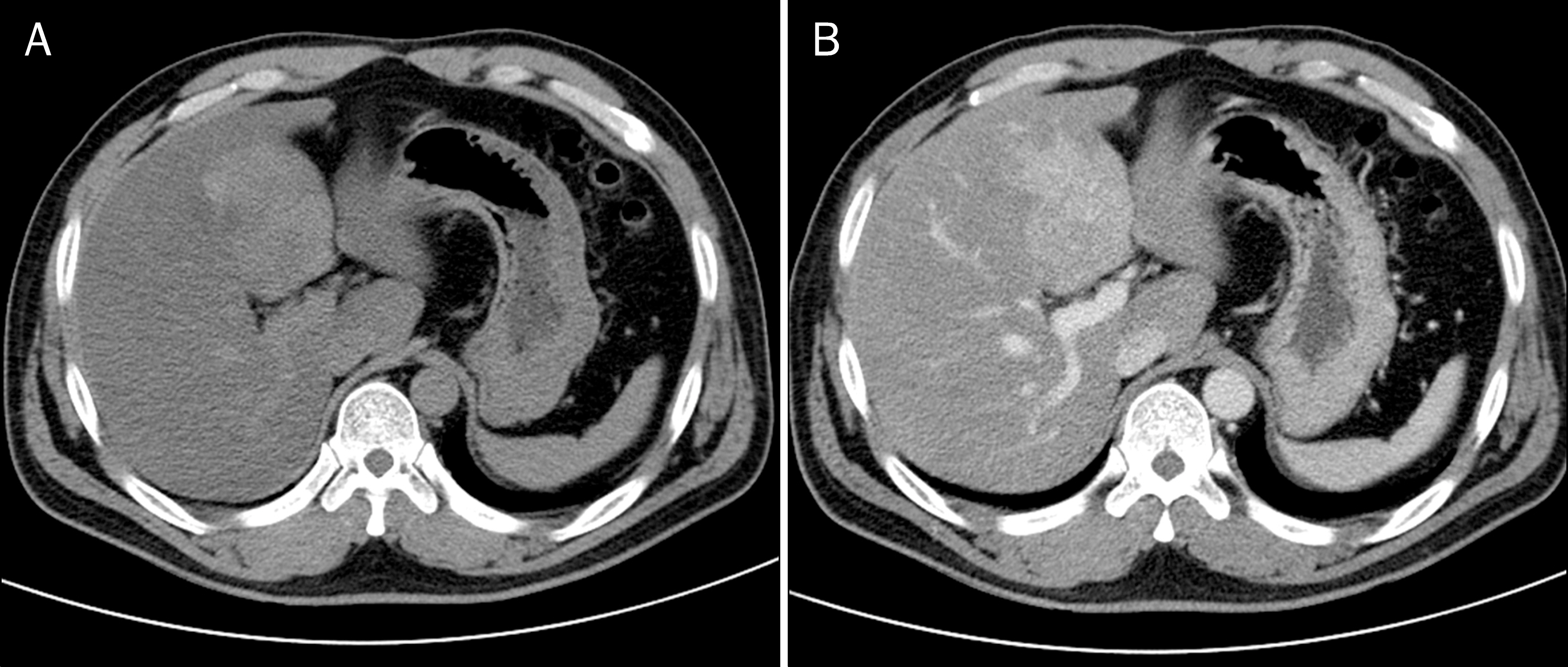
Fig. 2. Abdominal CT scan reveals 6.6×5.3 cm sized mass-like lesion with enhancing at both pre-enhance phase (A) and portal phase (B) images. Axial contrast enhanced portal phase image shows a diffusely hypodense liver (55 hounsfield units [HU]) in comparison with the spleen (110 HU) with a spared zone (segment IV).
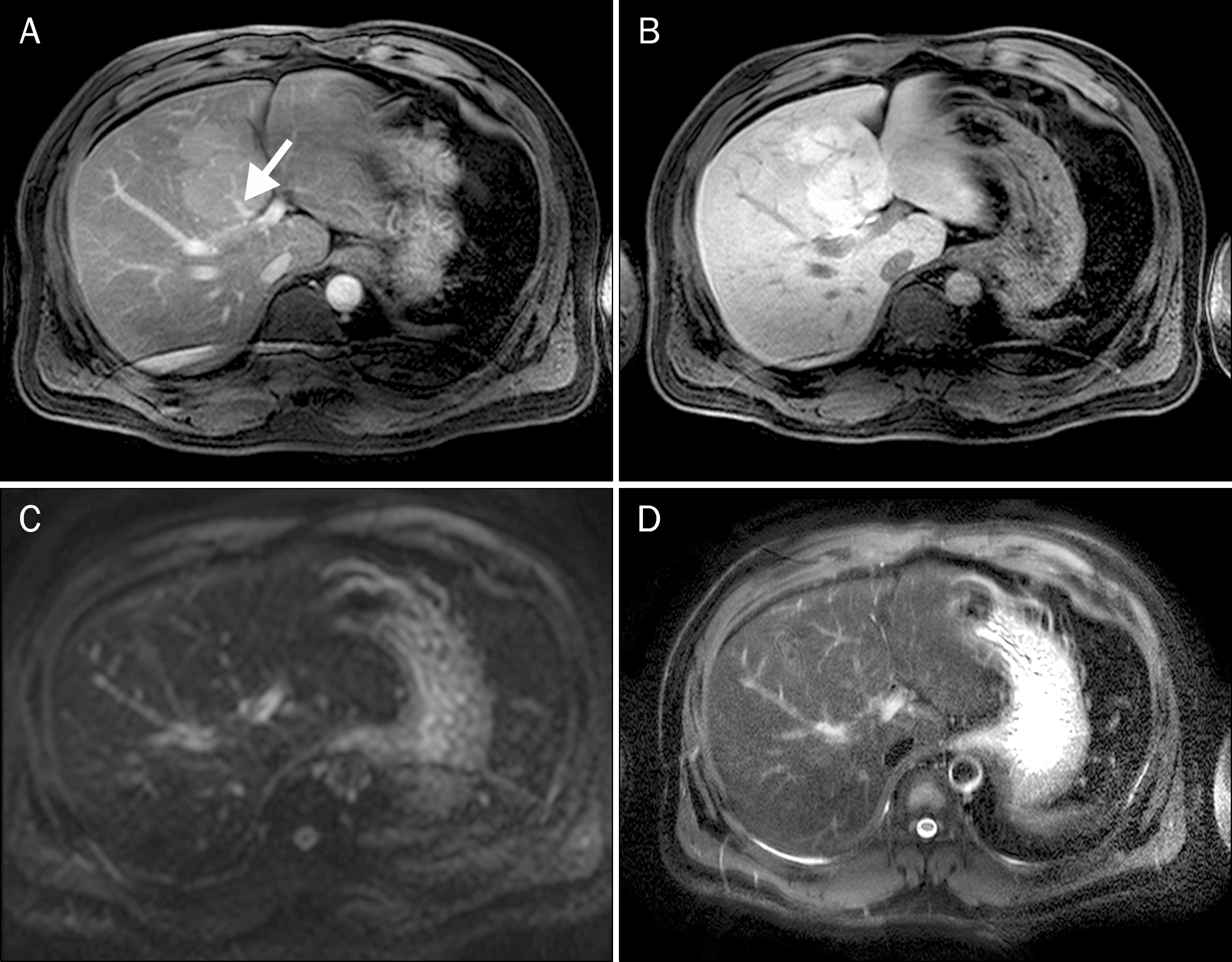
Fig. 3. Liver MRI. (A) Portal phase shows a lobulating, enhancing mass at segment IV of Liver. There is noted a aberrant right gastric vein in mass at segement IV of liver (arrow). (B) Hepatobiliary phase reveals high signal intensity at segment IV of liver. Diffusion weighted (C) and T2-weighted (D) images show no significant signal changes between segment IV of liver and the other part of liver.

Fig. 4. Liver MRI. Axial T1-weighted (A) in-phase and out of phase (B) images shows an important signal drop of the liver on the opposed-phase image with the exception of spared zone in segment IV of the liver.
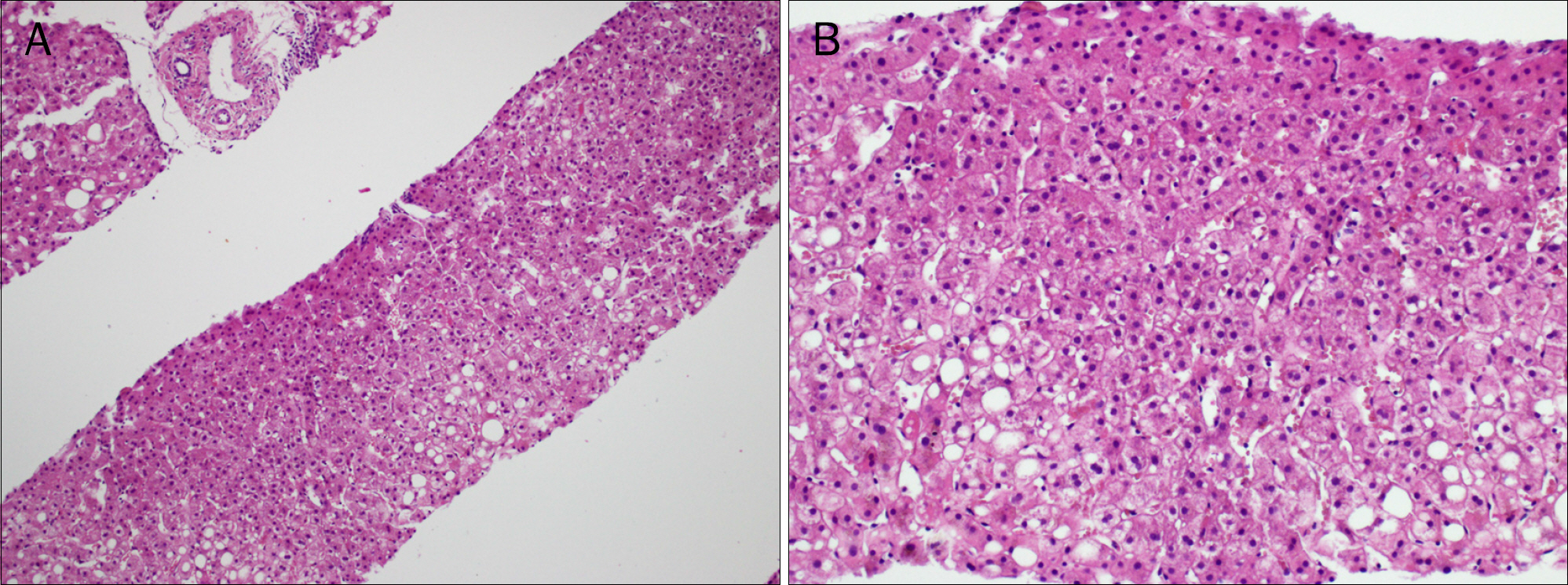
Fig. 5. Microscopic findings of liver (H&E). (A) Hepatic steatosis was observed but there were noted some regions without fatty changes (×100).(B) In addition to hepatic steatosis, balloon degeneration, periportal inflammation, perisinusoidal fibrosis were also present (×200).
Reference
-
References
1. Preiss D, Sattar N. Nonalcoholic fatty liver disease: an overview of prevalence, diagnosis, pathogenesis and treatment considerations. Clin Sci (Lond). 2008; 115:141–150.
Article2. Ministry of Food and Drug Safety. Influence of dietary intake on non-alcoholic fatty liver disease in Korean. Cheongwon: Ministry of Food and Drug Safety;2012.3. Boyce CJ, Pickhardt PJ, Kim DH, et al. Hepatic steatosis (fatty liver disease) in asymptomatic adults identified by unenhanced low-dose CT. AJR Am J Roentgenol. 2010; 194:623–628.
Article4. Alpern MB, Lawson TL, Foley WD, et al. Focal hepatic masses and fatty infiltration detected by enhanced dynamic CT. Radiology. 1986; 158:45–49.
Article5. Kratzer W, Akinli AS, Bommer M, et al. Prevalence and risk factors of focal sparing in hepatic steatosis. Ultraschall Med. 2010; 31:37–42.6. Kemper J, Jung G, Poll LW, Jonkmanns C, Lüthen R, Moedder U. CT and MRI findings of multifocal hepatic steatosis mimicking malignancy. Abdom Imaging. 2002; 27:708–710.
Article7. McKenzie A, Gill G, McIntosh R, Hennessy O, Pryde D. Computed tomographic and ultrasound appearances of focal spared areas in fatty infiltration of the liver. Australas Radiol. 1991; 35:166–168.
Article8. Chong VF, Fan YF. Ultrasonographic hepatic pseudolesions: normal parenchyma mimicking mass lesions in fatty liver. Clin Radiol. 1994; 49:326–329.
Article9. Hamer OW, Aguirre DA, Casola G, Lavine JE, Woenckhaus M, Sirlin CB. Fatty liver: imaging patterns and pitfalls. Radiographics. 2006; 26:1637–1653.
Article10. Basaran C, Karcaaltincaba M, Akata D, et al. Fat-containing lesions of the liver: cross-sectional imaging findings with emphasis on MRI. AJR Am J Roentgenol. 2005; 184:1103–1110.
Article11. Valls C, Iannacconne R, Alba E, et al. Fat in the liver: diagnosis and characterization. Eur Radiol. 2006; 16:2292–2308.
Article12. Mathieu D, Luciani A, Achab A, Zegai B, Bouanane M, Kobeiter H. Les pseudo-lésions hépatiques. Gastroenterol Clin Biol. 2001; 25:B158–B166.13. Matsui O, Kadoya M, Takahashi S, et al. Focal sparing of segment IV in fatty livers shown by sonography and CT: correlation with aberrant gastric venous drainage. AJR Am J Roentgenol. 1995; 164:1137–1140.
Article
- Full Text Links
-
- Actions
-
Cited
- CITED
-
- Close
- Share
-
- Similar articles
-
- Focal Sparing of the Fatty Liver Caused by a Nontumorous Arterioportal Shunt
- Focal Sparing in Fatty Liver: Mimicking Hypervascular Tumor on Gadolinium-Enhanced Opposed-Phase Gradient-Echo Images
- Fatty Liver
- Impact of variations in fatty liver on sonographic detection of focal hepatic lesions originally identified by CT
- Focal Fatty Change of the Liver
