A Newly Developed Stent Thrombus Related to Optical Coherence Tomography
- Affiliations
-
- 1Division of Cardiology, Yonsei Cardiovascular Center, Yonsei University College of Medicine, Seoul, Korea. kjs1218@yuhs.ac
- KMID: 2225702
- DOI: http://doi.org/10.4070/kcj.2008.38.12.674
Abstract
- Optical coherence tomography (OCT) is a useful coronary imaging tool for atherosclerotic plaque characterization and stent evaluation. However, proximal balloon inflation is required in order to reduce signal changes caused by red blood cells and to acquire a clear image. One rare acute complication related to proximal balloon occlusion is micro-thrombus formation. We report a case of multiple, acute micro-thrombi forming after an OCT procedure, despite the use of appropriate prevention for intracoronary thrombus formation.
Keyword
MeSH Terms
Figure
-
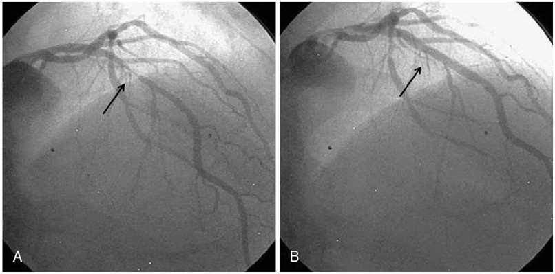
Fig. 1 Coronary angiography (CAG) before 9 months showed the culprit lesion (dark arrow) in the middle segment of the left anterior descending (LAD) artery (A), and CAG after zotarolimus-eluting stent implantation (B) showed that the stenosis of the culprit lesion (dark arrow) had resolved.
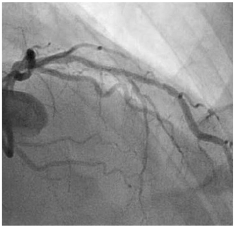
Fig. 2 Follow-up coronary angiography showed a patent stent in the middle segment of the left anterior descending (LAD) artery.
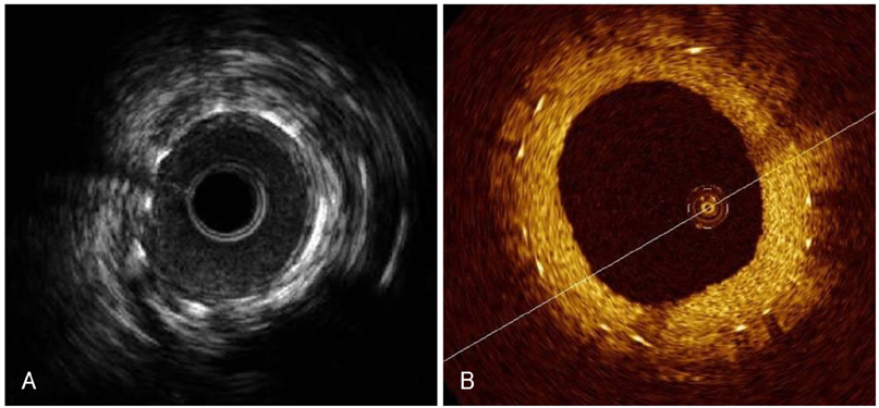
Fig. 3 Intravascular ultrasound (IVUS) (A) and optical coherence tomography (OCT) (B) showed the neointima covering the stent struts and no thrombus formation.

Fig. 4 Optical coherence tomography (OCT) revealed acute thrombus formation (white arrow) during the pull-back portion of the procedure (A). Follow-up coronary angiography after OCT showed total occlusion in the far distal segment of the left anterior descending (LAD) (dark arrow) (B) due to thromboembolism.
Reference
-
1. Bouma BE, Tearney GJ, Yabushita H, et al. Evaluation of intracoronary stenting by intravascular optical coherence tomography. Heart. 2003. 89:317–320.2. Kim BK, Oh SJ, Jeon DW, Kim KH, Yang JY. Is stent underexpansion the main cause of in-stent restenosis after sirolimus-eluting stent implantation?: an intravascular ultrasound study. Korean Circ J. 2007. 37:58–63.3. Hong YJ, Jeong MH, Ahn YK, et al. Effect of conventional dose of simvastatin on plaque regression and vascular remodeling in the peristent reference segments of normocholesterolemic patients: a serial intravascular ultrasound assessment. Korean Circ J. 2007. 37:483–488.4. Jang IK, Bouma BE, Kang DH, et al. Visualization of coronary atherosclerotic plaques in patients using optical coherence tomography: comparison with intravascular ultrasound. J Am Coll Cardiol. 2002. 39:604–609.5. Barlis P, Serruys PW, Devries A, Regar E. Optical coherence tomography assessment of vulnerable plaque rupture: predilection for the plaque 'shoulder'. Eur Heart J. 2008. 29:2023.6. Burke AP, Farb A, Kolodgie FD, Narula J, Virmani R. Atherosclerotic plaque morphology and coronary thrombi. J Nucl Cardiol. 2002. 9:95–103.7. Kume T, Akasaka T, Kawamoto T, et al. Assessment of coronary arterial plaque by optical coherence tomography. Am J Cardiol. 2006. 97:1172–1175.8. Stamper D, Weissman NJ, Brezinski M. Plaque characterization with optical coherence tomography. J Am Coll Cardiol. 2006. 47:8 Suppl. C69–C79.9. Brezinski ME, Tearney GJ, Weissman NJ, et al. Assessing atherosclerotic plaque morphology: comparison of optical coherence tomography and high frequency intravascular ultrasound. Heart. 1997. 77:397–403.10. Jang IK, Tearney GJ, MacNeill B, et al. In vivo characterization of coronary atherosclerotic plaque by use of optical coherence tomography. Circulation. 2005. 111:1551–1555.11. Pinto TL, Waksman R. Clinical applications of optical coherence tomography. J Interv Cardiol. 2006. 19:566–573.
- Full Text Links
-
- Actions
-
Cited
- CITED
-
- Close
- Share
-
- Similar articles
-
- A Newly Formed and Ruptured Atheromatous Plaque within Neointima after Drug-Eluting Stent Implantation: 2-Year Follow-Up Intravascular Ultrasound and Optical Coherence Tomography Studies
- Identification of Vulnerable Plaque in a Stented Coronary Segment 17 Years after Implantation Using Optical Coherence Tomography
- Availability of Optical Coherence Tomography in Diagnosis and Classification of Choroidal Neovascularization
- Optical Coherence Tomography and Stent Boost Imaging Guided Bioresorbable Vascular Scaffold Overlapping for Coronary Chronic Total Occlusion Lesion
- Utility of Optical Coherence Tomography to Assess a Hazy Intracoronary Image after Percutaneous Coronary Intervention
