Korean Circ J.
2009 Apr;39(4):145-150. 10.4070/kcj.2009.39.4.145.
The Influence of the Left Ventricular Geometry on the Left Atrial Size and Left Ventricular Filling Pressure in Hypertensive Patients, as Assessed by Echocardiography
- Affiliations
-
- 1Cardiovascular Center, Department of Internal Medicine, School of Medicine, Ewha Womans University, Seoul, Korea. giljshin@ewha.ac.kr
- KMID: 2225689
- DOI: http://doi.org/10.4070/kcj.2009.39.4.145
Abstract
- BACKGROUND AND OBJECTIVES
It is not well understood whether the left ventricular geometry is associated with such diastolic parameters as the left atrial volume and the left ventricular filling pressure, as assessed by the Doppler indices. Accordingly, this study aimed to evaluate the influence of the left ventricular geometry on the left atrial volume and the left ventricular filling pressure, as assessed by the Doppler indices. SUBJECTS AND METHODS: 181 patients (mean age: 63+/-9 years old, 62 males) with hypertension were included for echocardiographic analysis. The hypertensive patients were classified into four groups according to the left ventricular mass index and the relative wall thickness: normal geometry, concentric remodeling, eccentric hypertrophy and concentric hypertrophy. We excluded all the individuals with established cardiovascular disease, atrial fibrillation, significant aortic and/or mitral valve disease, or an ejection fraction <50%. RESULTS: By definition, the left ventricular mass was increased in the patients with eccentric and concentric hypertrophy. Both the left ventricular end-systolic diameter and the left ventricular end-diastolic diameter were reduced in the concentric remodeling group, whereas the left ventricular end-systolic diameter and the left ventricular end-diastolic diameter were increased in the eccentric and concentric hypertrophy groups. Compared with the patients with normal geometry, the patients with eccentric and concentric hypertrophy demonstrated a significant higher value for the left atrial volume index. The ratio of the transmitral inflow velocity to the mitral annular velocity (E/E') showed a stepwise increase from the patients with normal geometry to the patients with concentric remodeling, and then to the patients with eccentric and concentric hypertrophy. CONCLUSION: This study demonstrates that in a patient population with hypertension and who are without systolic dysfunction, the left atrial volume index and the E/E' demonstrated a progressive worsening of the left ventricular diastolic function from patients with normal geometry to the patients with concentric remodeling, and then to the patients with eccentric and concentric hypertrophy.
MeSH Terms
Figure
-

Fig. 1 Measurement of the mitral inflow velocities by pulsed-wave Doppler (A) and measurement of the mitral annular velocity by pulsed-wave Doppler tissue imaging (B). E: early diastolic mitral inflow velocity, A: late diastolic mitral inflow velocity, DT: deceleration time, E': early diastolic mitral annular tissue velocity, A': late diastolic mitral annular tissue velocity.
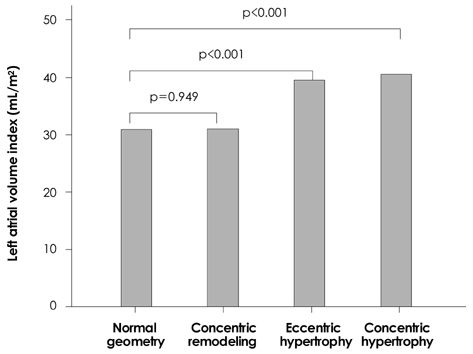
Fig. 2 The relationship of the left ventricular geometric pattern to the left atrial volume index.
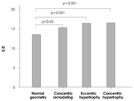
Fig. 3 The relationship of the left ventricular geometric pattern to the E/E'. E/E: the ratio of the transmitral inflow velocity to the mitral annular velocity.
Reference
-
1. Ganau A, Devereux RB, Roman MJ, et al. Patterns of left ventricular hypertrophy and geometric remodeling in essential hypertension. J Am Coll Cardiol. 1992. 19:1550–1558.2. Balci B, Yilmaz O. Influence of left ventricular geometry on regional systolic and diastolic function in patients with essential hypertension. Scand Cardiovasc J. 2002. 36:292–296.3. Devereux RB, James GD, Pickering TG. What is normal blood pressure?: comparison of ambulatory pressure level and variability in patients with normal or abnormal left ventricular geometry. Am J Hypertens. 1993. 6:211S–215S.4. Im SA, Jung HK, Park SH, Shin GJ, Lee WH. Patterns of left ventricular hypertrophy and geometric remodeling in essential hypertension. Korean Circ J. 1995. 25:423–433.5. Verdecchia P, Schillaci G, Borgioni C, et al. Adverse prognostic significance of concentric remodeling of the left ventricle in hypertensive subjects with normal left ventricular mass. J Am Coll Cardiol. 1995. 25:871–878.6. de Simone G, Kitzman DW, Chinali M, et al. Left ventricular concentric geometry is associated with impaired relaxation in hypertension. Eur Heart J. 2005. 26:1039–1045.7. Sahn DJ, Demaria A, Kisslo J, Weyman A. Recommendations regarding quantitation in M-mode echocardiography: results of a survey of echocardiographic measurements. Circulation. 1978. 58:1072–1083.8. Devereux RB, Alonso DR, Lutas EM, et al. Echocardiographic assessment of left ventricular hypertrophy: comparison to necropsy findings. Am J Cardiol. 1986. 57:450–458.9. Hammond LW, Devereux RB, Alderman MH, et al. The prevalence and correlates of echocardiographic left ventricular hypertrophy among employed patients with uncomplicated hypertension. J Am Coll Cardiol. 1986. 7:639–650.10. Pritchett AM, Jacobsen SJ, Mahoney DW, Rodeheffer RJ, Bailey KR, Redfield MM. Left atrial volume as an index of left atrial size: a population-based study. J Am Coll Cardiol. 2003. 41:1036–1043.11. Kim KS. The usefulness of Doppler tissue image in evaluation of left ventricular systolic and diastolic dysfunction. Korean Circ J. 2002. 32:99–105.12. Ommen SR, Nishimura RA, Appleton CP, et al. Clinical utility of Doppler echocardiography and tissue Doppler imaging in the estimation of left ventricular filling pressures: a comparative simultaneous Doppler-catheterization study. Circulation. 2000. 102:1788–1794.13. Lester SJ, Tajik AJ, Nishimura RA, Oh JK, Khandheria BK, Seward JB. Unlocking the mysteries of diastolic function: deciphering the Rosetta Stone 10 years later. J Am Coll Cardiol. 2008. 51:679–689.14. Miller JT, O'Rourke RA, Crawford MH. Left atrial enlargement: an early sign of hypertensive heart disease. Am Heart J. 1988. 116:1048–1051.15. Fouad FM, Slominsk JM, Tarazi RC. Left ventricular diastolic function in hypertension: relation to left ventricular mass and systolic function. J Am Coll Cardiol. 1984. 3:1500–1506.16. Douglas PS, Berko B, Lesh M, Reichk N. Alterations in diastolic function in response to progressive left ventricular hypertrophy. J Am Coll Cardiol. 1989. 13:461–467.17. Galderisi M, Caso P, Severino S, et al. Myocardial diastolic impairment caused by left ventricular hypertrophy involves basal septum more than other walls: analysis by pulsed Doppler tissue imaging. J Hypertens. 1999. 17:685–693.18. Marabotti C, Genovesi-Ebert A, Ghione S, Giaconi S, Palombo C. Diastolic function in the different patterns of left ventricular adaptation to essential hypertension. Int J Cardiol. 1994. 44:73–78.19. Verdecchia P, Schillaci G, Borgioni C, et al. Adverse prognostic significance of concentric remodeling of the left ventricle in hypertensive patients with normal left ventricular mass. J Am Coll Cardiol. 1995. 25:871–878.20. Pierdomenico SD, Lapenna D, Bucci A, Manente BM, Cuccurullo F, Mezzetti A. Prognostic value of left ventricular concentric remodeling in uncomplicated mild hypertension. Am J Hypertens. 2004. 17:1035–1039.
- Full Text Links
-
- Actions
-
Cited
- CITED
-
- Close
- Share
-
- Similar articles
-
- Evaluation of the Left Atrial Size and Function in Addition to Analysis of the Mitral and Pulmonary Venous Flow Velocity in the Estimation of Left Ventricular Filling Pressures
- Left Ventricular Diastolic Functions by M-Mode Echocardiogram in Essential Hypertensive Patients
- Interrlationship between Left Ventricular Mass and Diurnal Variations of Blood Pressure in Patients with Esssntial Hypertension
- The Changes of the Velocities of the Motions of the Posterior Aortic Wall in Hypertensive Heart Disease
- A Study for the Left Ventricular Diastolic Function in Mild to Moderate Hypertensive Patients without Left Ventricular Hypertrophy
