Slow Coronary Flow is Related to Increased Carotid Intima-Media Thickness but Not Pulse Wave Velocity
- Affiliations
-
- 1Department of Cardiovascular Medicine, Konkuk University School of Medicine, Seoul, Korea. khryumd@hanmail.net
- KMID: 2225075
- DOI: http://doi.org/10.4070/kcj.2011.41.11.666
Abstract
- BACKGROUND AND OBJECTIVES
Slow coronary flow (SCF) is characterized by delayed contrast dye opacification without significant stenosis of epicardial coronary arteries. However, the pathophysiology and clinical implications of SCF are not fully understood. Some reports have suggested that SCF might be caused by atherosclerosis in the coronary artery microvasculature. Measuring carotid intima-media thickness (IMT) and pulse wave velocity (PWV), which are non-invasive and simple diagnostic tools, was developed to detect subclinical atherosclerosis. Thus, we determined IMT and PWV, and their possible relationship in a SCF group and a normal coronary flow (NCF) group of patients.
SUBJECTS AND METHODS
We included 101 patients who complained of chest pain but had a normal coronary angiogram. Thrombolysis in Myocardial Infarction frame count (TIMI frame count, TFC) was evaluated in the left and right coronary arteries. We defined SCF as a TFC of more than 25. Carotid IMT was measured by ultrasonography in both common carotid arteries. PWV was calculated from pulse transit time between the brachial and ankle arteries.
RESULTS
Fifteen patients were included in the SCF group and 86 patients in the NCF group. Male patients (n=11, 73.3%) were significantly more common in the SCF group than in the NCF group (n=37, 43.0%, p<0.05). The TFC of the SCF and NCF groups were 28.8+/-3.5 and 15.7+/-4.5, respectively. The carotid IMT in the SCF group increased significantly compared to that in the NCF group (1.2+/-0.3 mm vs. 0.8+/-0.1 mm, p<0.01). However, no significant difference in PWV was observed between the two groups.
CONCLUSION
SCF may reflect early atherosclerotic changes in the coronary artery microvasculature.
MeSH Terms
Figure
-
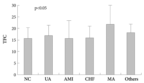
Fig. 1 TFC for each clinical diagnosis. TFC increased significantly in the microvascular angina group compared to other clinical diagnoses (p<0.05 by analysis of variance). AMI: acute myocardial infarction, CHF: chronic heart failure, MA: microvascular angina, NC: non-cardiac chest pain, TFC: Thrombolysis in Myocardial Infarction frame count, UA: unstable angina.

Fig. 2 A: a positive correlation was observed between carotid intima-media thickness (IMT) and the Thrombolysis in Myocardial Infarction frame count (TFC) (r=0.40, p<0.001). B: no significant correlation was found between pulse wave velocity (PWV) and the TFC. NS: not significant.
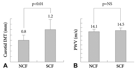
Fig. 3 Comparison of the carotid intima-media thickness (IMT) and pulse wave velocity (PWV) between the normal coronary flow (NCF) and slow coronary flow (SCF) groups. A: carotid IMT increased significantly in the SCF group compared to that in the NCF group. B: no difference in the PWV between the SCF and NCF groups. NS: not significant.
Reference
-
1. Beltrame JF, Limaye SB, Horowitz JD. The coronary slow flow phenomenon: a new coronary microvascular disorder. Cardiology. 2002. 97:197–202.2. Tambe AA, Demany MA, Zimmerman HA, Mascarenhas E. Angina pectoris and slow flow velocity of dye in coronary arteries: a new angiographic finding. Am Heart J. 1972. 84:66–71.3. Vrints C, Herman AG. Role of the endothelium in the regulation of coronary artery tone. Acta Cardiol. 1991. 46:399–418.4. Kurtoglu N, Akcay A, Dindar I. Usefulness of oral dipyridamole therapy for angiographic slow coronary artery flow. Am J Cardiol. 2001. 87:777–779.5. Motz W, Vogt M, Rabenau O, Scheler S, Lückhoff A, Strauer BE. Evidence of endothelial dysfunction in coronary resistance vessels, in patients with angina pectoris and normal coronary angiograms. Am J Cardiol. 1991. 68:996–1003.6. Quyyumi AA, Cannon RO 3rd, Panza JA, Diodati JG, Epstein SE. Endothelial dysfunction in patients with chest pain and normal coronary arteries. Circulation. 1992. 86:1864–1871.7. Egashira K, Inou T, Hirooka Y, Yamada A, Urabe Y, Takeshita A. Evidence of impaired endothelium dependent coronary vasodilatation in patients with angina pectoris and normal coronary angiograms. N Engl J Med. 1993. 328:1659–1664.8. Zeiher AM, Krause T, Schächinger V, Minners J, Moser E. Impaired endothelium: dependent vasodilation of the coronary resistance vessels is associated with exercise-induced myocardial ischemia. Circulation. 1995. 91:2345–2352.9. Camsari A, Ozcan T, Ozer C, Akcay B. Carotid artery intima-media thickness correlates with intravascular ultrasound parameters in patients with slow coronary flow. Atherosclerosis. 2008. 200:310–314.10. Wofford JL, Kahl FR, Howard GR, McKinney WM, Toole JF, Crouse JR 3rd. Relation of extent of extracranial carotid artery atherosclerosis as measured by B-mode ultrasound to the extent of coronary atherosclerosis. Arterioscler Thromb. 1991. 11:1786–1794.11. Craven TE, Ryu JE, Espeland MA, et al. Evaluation of the associations between carotid artery atherosclerosis and coronary artery stenosis: a case-control study. Circulation. 1990. 82:1230–1242.12. van Popele NM, Grobbee DE, Bots ML, et al. Association between arterial stiffness and atherosclerosis: the Rotterdam Study. Stroke. 2001. 32:454–460.13. Gibson CM, Cannon CP, Daley WL, et al. TIMI frame count: a quantitative method of assessing coronary artery flow. Circulation. 1996. 93:879–888.14. Tanriverdi H, Evrengul H, Tanriverdi S, et al. Carotid intima-media thickness in coronary slow flow: relationship with plasma homocysteine levels. Coron Artery Dis. 2006. 17:331–337.15. Yamashina A, Tomiyama H, Takeda K, et al. Validity, reproducibility, and clinical significance of noninvasive brachial-ankle pulse wave velocity measurement. Hypertens Res. 2002. 25:359–364.16. Saya S, Hennebry TA, Lozano P, Lazzara R, Schechter E. Coronary slow flow phenomenon and risk for sudden cardiac death due to ventricular arrhythmias: a case report and review of literature. Clin Cardiol. 2008. 31:352–355.17. Olivotto I, Cecchi F, Gistri R, et al. Relevance of coronary microvascular flow impairment to long-term remodeling and systolic dysfunction in hypertrophic cardiomyopathy. J Am Coll Cardiol. 2006. 47:1043–1048.18. Cin VG, Pekdemir H, Camsar A, et al. Diffuse intimal thickening of coronary arteries in slow coronary flow. Jpn Heart J. 2003. 44:907–919.19. Cakmak M, Tanriverdi H, Cakmak N, Evrengul H, Cetemen S, Kuru O. Simvastatin may improve myocardial perfusion abnormality in slow coronary flow. Cardiology. 2008. 110:39–44.20. Yigit F, Sezgin AT, Demircan S, Tekin G, Erol T, Muderrisoglu H. Slow coronary flow is associated with carotid artery dilatation. Tohoku J Exp Med. 2006. 209:41–48.21. Matsushima Y, Kawano H, Koide Y, et al. Relationship of carotid intima-media thickness, pulse wave velocity, and ankle brachial index to the severity of coronary artery atherosclerosis. Clin Cardiol. 2004. 27:629–634.22. Megnien JL, Simon A, Denarie N, et al. Aortic stiffening does not predict coronary and extracoronary atherosclerosis in asymptomatic men at risk for cardiovascular disease. Am J Hypertens. 1998. 11:293–301.23. Seo WW, Chang HJ, Cho I, et al. The value of brachial-ankle pulse wave velocity as a predictor of coronary artery disease in high-risk patients. Korean Circ J. 2010. 40:224–229. doi:10.4070/kcj.2010.40.5.224.
- Full Text Links
-
- Actions
-
Cited
- CITED
-
- Close
- Share
-
- Similar articles
-
- The Relationship of Pulse-wave Velocity with Carotid Intima-media Thickness and Carotid Artery Distensibility
- Carotid ultrasound in patients with coronary artery disease
- Impacts of Atherosclerotic Coronary Risk Factors on Atherosclerotic Surrogates in Patients with Coronary Artery Disease
- Noninvasive surrogates in assessing the severity of coronary atherosclerosis
- Intima-Media Thickness and Pulse Wave Velocity in Hypertensive Adolescents
