Korean Circ J.
2012 Mar;42(3):205-207. 10.4070/kcj.2012.42.3.205.
Contrast Echo-A Simple Diagnostic Tool for a Coronary Artery Fistula
- Affiliations
-
- 1Cardiology Division, National Health Insurance Corporation Ilsan Hospital, Goyang, Korea. drpuooh@hanmail.net
- 2Cardiology Division, Gangnam Severance Hospital, Yonsei University College of Medicine, Seoul, Korea.
- KMID: 2225036
- DOI: http://doi.org/10.4070/kcj.2012.42.3.205
Abstract
- Coronary artery fistulas have been diagnosed with aortography, coronary angiography, and coronary computed tomography (CT). A large fistula can be occasionally found as a mass lesion on echocardiography but cannot be easily confirmed. Here, we report a new diagnostic approach to coronary artery fistulas using a contrast agent and transthoracic echocardiography. Transthoracic echocardiography of a 46-year-old female suffering from dyspnea revealed suspicious small turbulent flow in the main pulmonary artery. Following infusion of a contrast agent, we found whitish flow in the main pulmonary artery during the diastolic phase, and aortic CT revealed two huge right coronary artery fistulas in the main pulmonary artery. A simple diagnostic approach to a coronary artery fistula using contrast agent helped us confirm the diagnosis because of the typical diastolic whitish flow in the pulmonary artery.
MeSH Terms
Figure
-

Fig. 1 Echocardiographic findings of a coronary fistula with Doppler and contrast agent. Parasternal short axis view showing abnormal, small turbulent flow in the main pulmonary artery (A), and Doppler revealed diastolic-dominant flow (B, white arrow heads). Systolic and diastolic flow in the pulmonary artery were shown with contrast infusion (C and D), and an unusual whitish flow was noted in the main pulmonary artery during the diastolic phase (D, white arrow heads).
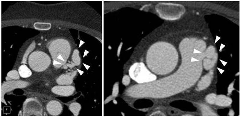
Fig. 2 Computed tomographic findings of the coronary fistula. Aortic computed tomography revealed two huge right coronary artery fistulas in the main pulmonary artery (white arrows).
Reference
-
1. Wei K, Mulvagh SL, Carson L, et al. The safety of definity and optison for ultrasound image enhancement: a retrospective analysis of 78,383 administered contrast doses. J Am Soc Echocardiogr. 2008. 21:1202–1206.2. Burch GH, Sahn DJ. Congenital coronary artery anomalies: the pediatric perspective. Coron Artery Dis. 2001. 12:605–616.3. Luo L, Kebede S, Wu S, Stouffer GA. Coronary artery fistulae. Am J Med Sci. 2006. 332:79–84.4. Morgan JR, Forker AD, O'Sullivan MJ Jr, Fosburg RG. Coronary arterial fistulas: seven cases with unusual features. Am J Cardiol. 1972. 30:432–436.5. Dresios C, Apostolakis S, Tzortzis S, Lazaridis K, Gardikiotis A. Apical hypertrophic cardiomyopathy associated with multiple coronary artery-left ventricular fistulae: a report of a case and review of the literature. Eur J Echocardiogr. 2010. 11:E9.6. Yamanaka O, Hobbs RE. Coronary artery anomalies in 126,595 patients undergoing coronary arteriography. Cathet Cardiovasc Diagn. 1990. 21:28–40.7. Zenooz NA, Habibi R, Mammen L, Finn JP, Gilkeson RC. Coronary artery fistulas: CT findings. Radiographics. 2009. 29:781–789.8. Kim YH, Lee SH, Kang CH, et al. A case report of coronary arteriovenous fistula. Korean Circ J. 1982. 12:189–192.9. Lee SJ, Her SH, Jin SW, et al. A case of bilateral coronary to pulmonary artery fistulas associated with severe aortic regurgitation. Korean Circ J. 2008. 38:331–334.10. Choi SH, Seo HS, Oh SJ, et al. A case of multiple coronary artery-left ventricular microfistulae demonstrated by transthoracic Doppler echocardiography. Korean Circ J. 2003. 33:338–342.
- Full Text Links
-
- Actions
-
Cited
- CITED
-
- Close
- Share
-
- Similar articles
-
- A case of coronary artery-pulmonary artery fistula communicated with aorto-pulmonary fistula via common channel detected by Multidetector row CT (MDCT) and coronary angiography
- Three Cases of Coronary Artery Fistula from Right Coronay to Left Ventricle
- Congenital Giant Left Circumflex Artery-to-Left Ventricle Fistula Detected Using Two-Dimensional and Doppler Echocardiography
- A Case of Double Right Coronary Artery with Arteriovenous Fisula
- A case report of coronary artery fistula to the left ventricle
