Coronary Slow Flow Phenomenon Leads to ST Elevation Myocardial Infarction
- Affiliations
-
- 1Department of Cardiology, Kutahya Evliya Celebi Education and Research Hospital, Kutahya, Turkey. medicineman_tr@hotmail.com
- KMID: 2224966
- DOI: http://doi.org/10.4070/kcj.2013.43.3.196
Abstract
- The exact etiology of the coronary slow flow phenomenon (CSFP) is not certain. CSFP is not a normal variant as it is an absolutely pathological entity. Furthermore, CSFP not only leads to myocardial ischemia but it can also cause classical acute ST elevation myocardial infarction, which necessitates coronary angiography for a definite diagnosis.
MeSH Terms
Figure
-
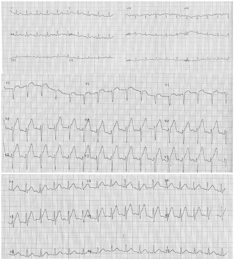
Fig. 1 The ECG shows marked ST elevation in the anterior precordial leads. This ECG leads to a diagnosis of hyperacute anterior myocardial infarction. ECG: electrocardiography.

Fig. 2 This angiographical image (A: left anterior oblique cranial view, B: right anterior oblique view) reveals left sided normal coronary arteries except slow coronary flow in the left anterior descending artery.

Fig. 3 The Thrombolysis in Myocardial Infarction (TIMI) frame count for the left anterior descending coronary artery (A and B) was 67-9=58. The corrected TIMI frame count was calculated as 58/1.7=34.1 (normal range 21±3).
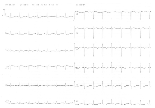
Fig. 4 After the chest pain ceased, the electrocardiography showed the resolution ST elevation and negative T waves between V 3-6 leads.
Reference
-
1. Mangieri E, Macchiarelli G, Ciavolella M, et al. Slow coronary flow: clinical and histopathological features in patients with otherwise normal epicardial coronary arteries. Cathet Cardiovasc Diagn. 1996. 37:375–381.2. Mosseri M, Yarom R, Gotsman MS, Hasin Y. Histologic evidence for small-vessel coronary artery disease in patients with angina pectoris and patent large coronary arteries. Circulation. 1986. 74:964–972.3. Singh S, Kothari SS, Bahl VK. Coronary slow flow phenomenon: an angiographic curiosity. Indian Heart J. 2004. 56:613–617.4. Gibson CM, Cannon CP, Daley WL, et al. TIMI frame count: a quantitative method of assessing coronary artery flow. Circulation. 1996. 93:879–888.5. Demirkol MO, Yaymaci B, Mutlu B. Dipyridamole myocardial perfusion single photon emission computed tomography in patients with slow coronary flow. Coron Artery Dis. 2002. 13:223–229.6. Kapoor A, Goel PK, Gupta S. Slow coronary flow--a cause for angina with ST segment elevation and normal coronary arteries. A case report. Int J Cardiol. 1998. 67:257–261.7. Tatli E, Yildirim T, Aktoz M. Does coronary slow flow phenomenon lead to myocardial ischemia? Int J Cardiol. 2009. 131:e101–e102.
- Full Text Links
-
- Actions
-
Cited
- CITED
-
- Close
- Share
-
- Similar articles
-
- Acute Myocardial Infarction by Right Coronary Artery Occlusion Presenting as Precordial ST Elevation on Electrocardiography
- Evolution of Diastolic Dysfunction in Patients with Coronary Slow Flow Phenomenon and Acute Non-ST Segment Elevation Myocardial Infarction
- Coronary Flow Doppler Profile in No-Reflex Phenomenon after Direct PTCA in Acute Myocardial Infarction
- Precordial ST-Segment Elevation in Acute Right Ventricular Myocardial Infarction
- A Case of Apical Hypertrophic Cardiomyopathy Combined Acute Myocardial Infarction with Multiple Coronary Thrombosis
