A Case of Partial Congenital Pericardial Defect Presenting as Acute Coronary Syndrome
- Affiliations
-
- 1Division of Internal Medicine, Sejong General Hospital, Bucheon, Korea. yoorimbin@sejongh.co.kr
- KMID: 2224783
- DOI: http://doi.org/10.4070/kcj.2013.43.12.845
Abstract
- Congenital pericardial defects are rare and asymptomatic for both partial and complete defects. However, some patients can experience syncope, arrhythmia, and chest pain. When a patient experiences a symptom, it may be caused by herniation and dynamic compression or torsion of a heart structure including the coronary arteries. Diagnosis of a congenital pericardial defect may be difficult, especially in old patients with concomitant coronary artery disease. The clinical importance of congenital pericardial defect has not been stressed and congenital pericardial defects are regarded as benign, but in this case, pericardial defect was responsible for myocardial ischemia. The authors report a case of partial congenital pericardial defect causing herniation and dynamic compression of the coronary arteries, presenting as an acute coronary syndrome in an old man, with an emphasis on the unique features of the coronary angiogram that support the diagnosis of partial pericardial defects.
MeSH Terms
Figure
-
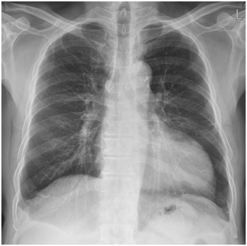
Fig. 1 Chest radiograph. Chest posteroanterior view shows levoposition of heart with a bulging of the left ventricle.
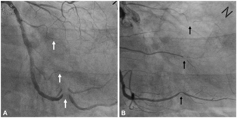
Fig. 2 Diagnostic coronary angiography. Coronary angiogram shows geographically circumferential phasic diastolic external compression of the obtuse marginal branches of the left circumflex artery (white arrows), right ventricular branches and the posterior descending artery of the right coronary artery (black arrows). A: left anterior oblique-caudal view. B: right anterior oblique-caudal view.
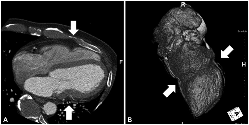
Fig. 3 Computed tomography of the heart. Cardiac CT shows a circumferential groove of the heart caused by external compression and impingement of the coronary arteries (arrows). A: cross sectional view. B: three-dimensional reconstruction view.

Fig. 4 The operative findings. The operative findings shows a rolled up thickened circumferential pericardial edge and the epicardial groove created by the dense fibrous rim of the pericardium just around the mid-level of the left ventricle.
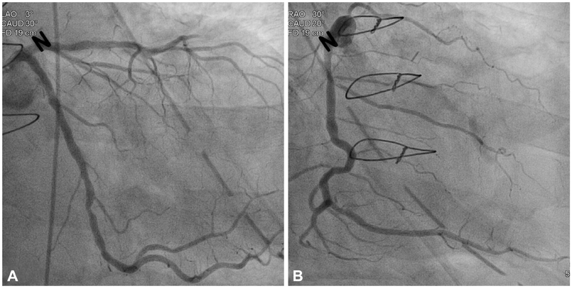
Fig. 5 A follow-up coronary angiography. Coronary angiogram show no features of the previous dynamic occlusion of the coronary arteries which were shown in Fig. 2. A: left anterior oblique-caudal view. B: right anterior oblique-caudal view.
Reference
-
1. Rusk RA, Kenny A. Congenital pericardial defect presenting as chest pain. Heart. 1999; 81:327–328.2. Kojima S, Nakamura T, Sugiyama S, et al. Cardiac displacement with a congenital complete left-sided pericardial defect in a patient with exertional angina pectoris. Angiology. 2004; 55:445–449.3. Fisher FD, Ehrenhaft JL. Congenital pericardial defect. JAMA. 1964; 188:78–81.4. Amiri A, Weber C, Schlosser V, Meinertz TH. Coronary artery disease in a patient with a congenital pericardial defect. Thorac Cardiovasc Surg. 1989; 37:379–381.5. Nguyen DQ, Wilson RF, Bolman RM 3rd, Park SJ. Congenital pericardial defect and concomitant coronary artery disease. Ann Thorac Surg. 2001; 72:1371–1373.6. Connolly HM, Click RL, Schattenberg TT, Seward JB, Tajik AJ. Congenital absence of the pericardium: echocardiography as a diagnostic tool. J Am Soc Echocardiogr. 1995; 8:87–92.7. Spodik D. Congenital abnormalities of the pericardium. The Pericardium: A Comprehensive Review. 1st ed. New York: Marcel Dekker;1997. p. 65–75.8. Spodik D. Pericardial diseases. In : Braunwald E, Zipes DP, Libby P, editors. Heart Disease: A Textbook of Cardiovascular Medicine. 6th ed. Philadelphia: Saunders;2001. p. 1823–1876.
