Ann Dermatol.
2009 May;21(2):171-173. 10.5021/ad.2009.21.2.171.
Giant Acral Melanoma on the Left Thumb of a Korean Patient
- Affiliations
-
- 1Department of Dermatology, College of Medicine, Korea University, Seoul, Korea. kumcihk@korea.ac.kr
- KMID: 2219389
- DOI: http://doi.org/10.5021/ad.2009.21.2.171
Abstract
- The acral regions of the limbs of Asians are predisposed to develop malignant melanoma, but giant-sized acral melanoma has not been previously reported in the Asian population. Giant-sized melanoma implies aggressive tumor invasion and so it is more difficult to achieve a therapeutic cure. A 56-year-old woman presented with a giant acral melanoma of the left thumb with concomitant bone destruction and axillary lymph node metastasis. The initial lesion was a subungual black macule on the left thumb that had grown into a giant 7.0*4.0*3.5 cm-sized melanoma over a 3 year period. The left thumb was amputated and the axillary lymph nodes were completely dissected. During the ensuing 3 months, she underwent adjuvant treatment with interferon-alpha-2a. The interesting feature of this case is that the large melanoma mass of this patient, which was accompanied with adjacent bone destruction and lymph node metastasis, had developed rapidly from a small black macule in the nail matrix, and this black macule was suspected to be a subungual melanoma.
MeSH Terms
Figure
-
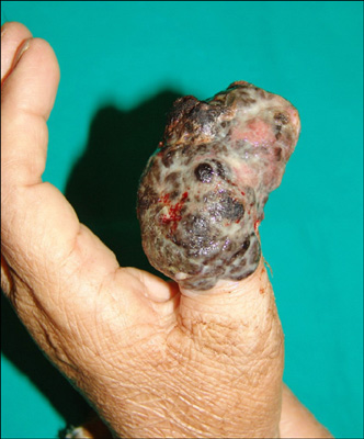
Fig. 1 A melanoma measuring 7.0×4.0×3.5 cm is surrounding the left thumb. Ulceration and spontaneous bleeding can be observed.
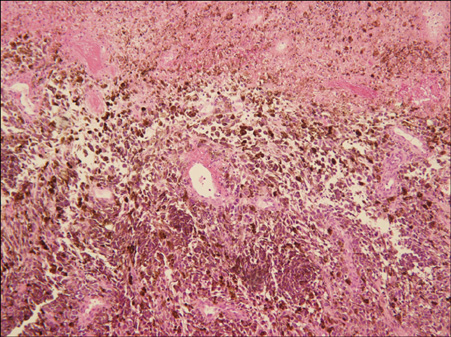
Fig. 2 Atypical epithelioid melanocytes with heavy melanin pigmentation are present at the dermo-epidermal junction and in the dermis (H&E, ×100).

Fig. 3 The distal phalangeal bone has largely been destroyed. There are also signs of tumor invasion on the dorsal radial aspect of the proximal phalanx. (A) X-ray image. (B) MRI image.
Reference
-
1. Benmeir P, Neuman A, Weinberg A, Sucher E, Weshler Z, Lusthaus S, et al. Giant melanoma of the inner thigh: a homeopathic life-threatening negligence. Ann Plast Surg. 1991. 27:583–585.
Article2. Pai RR, Kini H, Kamath SG, Kumar S. Giant hanging melanoma of the eyelid skin. Indian J Ophthalmol. 2008. 56:239–240.
Article3. Panajotovic L, Dordevic B, Pavlovic MD. A giant primary cutaneous melanoma of the scalp--can it be that big? J Eur Acad Dermatol Venereol. 2007. 21:1417–1418.4. Marano SR, Brooks RA, Spetzler RF, Rekate HL. Giant congenital cellular blue nevus of the scalp of a newborn with an underlying skull defect and invasion of the dura mater. Neurosurgery. 1986. 18:85–89.
Article5. Schneiderman H, Wu AY, Campbell WA, Forouhar F, Yamase H, Greenstein R, et al. Congenital melanoma with multiple prenatal metastases. Cancer. 1987. 60:1371–1377.
Article6. Morita K, Kudo H, Fujii K, Okamoto H, Matsubara K, Kanauchi H, et al. Giant metastatic malignant melanoma with an unknown primary site. J Dermatol. 1994. 21:442–446.
Article7. Zeebregts CJ, Schraffordt Koops H. Giant melanoma of the left thumb. Eur J Surg Oncol. 2000. 26:189–190.
Article8. De Giorgi V, Massi D, Carli P. Giant melanoma displaying gross features reproducing parameters seen on dermoscopy. Dermatol Surg. 2002. 28:646–647.
Article9. Enomoto T, Hamada M. Cryosurgery and OK 432 in the treatment of malignant melanoma. Arch Otolaryngol. 1984. 110:127–129.
Article10. Malik KP, Dadeya S, Gulliani BP, Gupta VS. Favourable outcome of giant malignant melanoma of the conjunctiva despite poor prognostic features. Can J Ophthalmol. 2003. 38:397–400.
Article
- Full Text Links
-
- Actions
-
Cited
- CITED
-
- Close
- Share
-
- Similar articles
-
- Acral Lentiginous Melanoma Developing during Long-standing Atypical Melanosis: Usefulness of Dermoscopy for Detection of Early Acral Melanoma
- A Case of Acral Lentiginous Melanoma
- Subungual Melanoma of Left Thumb: A Case Report
- The Appearance of a Candidate Site for a Primary Melanoma: A 5 Year-gap with a Melanoma of an Unknown Site
- Atypical Presentation of Subungal Melanoma
