J Korean Ophthalmol Soc.
2013 Jul;54(7):1139-1143. 10.3341/jkos.2013.54.7.1139.
Central Serous Chorioretinopathy in a Patient with Retinal Macrovessel
- Affiliations
-
- 1Department of Ophthalmology, Korea University College of Medicine, Seoul, Korea. ojr4991@yahoo.co.kr
- KMID: 2217430
- DOI: http://doi.org/10.3341/jkos.2013.54.7.1139
Abstract
- PURPOSE
The relationship between central serous chorioretinopathy associated with retinal macrovessel remains controversial due to its rareness. We report a case of central serous chorioretinopathy diagnosed by spectral domain optical coherence tomography (SD-OCT) in a patient with a retinal macrovessel that improved spontaneously.
CASE SUMMARY
A 36-year-old healthy male patient visited our clinic complaining of blurred vision in his left eye. Fundus examination of the left eye revealed central serous chorioretinopathy with retinal macrovessel in the macular area. Fundus fluorescent angiography showed an ink blot-shaped leakage, which was not clearly distinguishable due to a retinal macrovessel. Serous retinal detachment under the neurosensory retina was identified on OCT. A small pigment epithelial detachment was observed and considered as a leaking point. However, no visible exudates appeared to be leaking around the retinal macrovessel.
CONCLUSIONS
Central serous chorioretinopathy with a retinal macrovessel in a Korean patient was evaluated by SD-OCT. When compared with typical cases, no differences were observed in this case and no significant associations between central serous chorioretinopathy and retinal macrovessel were shown.
Keyword
MeSH Terms
Figure
-
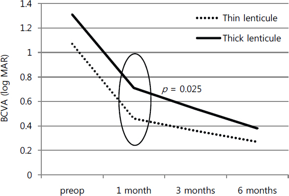
Figure 1. The comparison of visual prognosis after DSAEK surgery between thin lenticule group and thick lenticule group.
Reference
-
References
1. Lee WB, Jacobs DS, Musch DC, et al. Descemet's stripping endothelial keratoplasty: safety and outcomes: a report by the American Academy of Ophthalmology. Ophthalmology. 2009; 116:1818–30.2. Terry MA, Li J, Goshe J, Davis-Boozer D.Endothelial keratoplasty: the relationship between donor tissue size and donor endothelial survival. Ophthalmology. 2011; 118:1944–9.
Article3. Scorcia V, Matteoni S, Scorcia GB, et al. Pentacam assessment of posterior lamellar grafts to explain hyperopization after Descemet's stripping automated endothelial keratoplasty. Ophthalmology. 2009; 116:1651–5.
Article4. Pogorelov P, Cursiefen C, Bachmann BO, Kruse FE.Changes in donor corneal lenticule thickness after Descemet's stripping automated endothelial keratoplasty (DSAEK) with organ-cultured corneas. Br J Ophthalmol. 2009; 93:825–9.
Article5. Dapena I, Ham L, Melles GR.Endothelial keratoplasty: DSEK/ DSAEK or DMEK–the thinner the better? Curr Opin Ophthalmol. 2009; 20:299–307.6. Price MO, Giebel AW, Fairchild KM, Price FW Jr.Descemet's membrane endothelial keratoplasty: prospective multicenter study of visual and refractive outcomes and endothelial survival. Ophthalmology. 2009; 116:2361–8.7. McCauley MB, Price MO, Fairchild KM, et al. Prospective study of visual outcomes and endothelial survival with Descemet membrane automated endothelial keratoplasty. Cornea. 2011; 30:315–9.
Article8. Price FW Jr, Price MO.Descemet's stripping with endothelial keratoplasty in 50 eyes: a refractive neutral corneal transplant. J Refract Surg. 2005; 21:339–45.
Article9. Covert DJ, Koenig SB.Descemet stripping and automated endothelial keratoplasty (DSAEK) in eyes with failed penetrating keratoplasty. Cornea. 2007; 26:692–6.
Article10. Price MO, Price FW Jr.Descemet's stripping with endothelial keratoplasty: comparative outcomes with microkeratome-dissected and manually dissected donor tissue. Ophthalmology. 2006; 113:1936–42.11. Chen ES, Terry MA, Shamie N, et al. Endothelial keratoplasty: vision, endothelial survival, and complications in a comparative case series of fellows vs attending surgeons. Am J Ophthalmol. 2009; 148:26–31.e2.
Article12. Price MO, Gorovoy M, Benetz BA, et al. Descemet's stripping automated endothelial keratoplasty outcomes compared with penetrating keratoplasty from the Cornea Donor Study. Ophthalmology. 2010; 117:438–44.
Article13. Bahar I, Kaiserman I, Livny E, Slomovic A.Changes in corneal curvatures and anterior segment parameters after descemet stripping automated endothelial keratoplasty. Curr Eye Res. 2010; 35:961–6.
Article14. Lombardo M, Terry MA, Lombardo G, et al. Analysis of posterior donor corneal parameters 1 year after Descemet stripping automated endothelial keratoplasty (DSAEK) triple procedure. Graefes Arch Clin Exp Ophthalmol. 2010; 248:421–7.
Article15. Neff KD, Biber JM, Holland EJ.Comparison of central corneal graft thickness to visual acuity outcomes in endothelial keratoplasty. Cornea. 2011; 30:388–91.
Article16. Chen ES, Terry MA, Shamie N, et al. Precut tissue in Descemet's stripping automated endothelial keratoplasty donor characteristics and early postoperative complications. Ophthalmology. 2008; 115:497–502.17. Terry MA, Shamie N, Chen ES, et al. Precut tissue for Descemet's stripping automated endothelial keratoplasty: vision, astigmatism, and endothelial survival. Ophthalmology. 2009; 116:248–56.
- Full Text Links
-
- Actions
-
Cited
- CITED
-
- Close
- Share
-
- Similar articles
-
- A case of Atypical Central Serous Chorioretinopathy with Bullous Retinal Detachment
- Stellate Ganglion Block for Treatment of Central Serous Chorioretinopathy
- A Case of Atypical Idiopathic Central Serous Chorioretinopathy
- Measurement and Analysis of Neurosensory Retinal Detachment in Central Serous Chorioretinopathy Using Heidelberg Retina Tomograph
- A Case of Central Serous Chorioretinopathy with Bullous Retinal Detachment
