J Korean Ophthalmol Soc.
2013 Jul;54(7):1066-1073. 10.3341/jkos.2013.54.7.1066.
Changes of Peripapillary Retinal Nerve Fiber Layer Thickness Profile According to Aging in Myopic Eyes
- Affiliations
-
- 1Department of Ophthalmology, Kangdong Sacred Heart Hospital, Hallym University College of Medicine, Seoul, Korea. demian7435@gmail.com
- KMID: 2217417
- DOI: http://doi.org/10.3341/jkos.2013.54.7.1066
Abstract
- PURPOSE
To evaluate the effects of age on the distributional variability of peripapillary retinal nerve fiber layer (RFNL) thickness measured by optical coherence tomography (OCT) in myopia.
METHODS
Only the right eye of 64 myopic patients with long axial length (> or =24.5 mm) was included in the present study. The patients were divided into 2 age groups, 20 to 39 years of age and 40 to 59 years of age. Eventually, 42 subjects were selected and matched based on the difference of axial length not exceeding 0.5 mm between subjects in each group. The RFNL thickness was measured using Stratus OCT and average thickness, angular locations of double humps, and false-positive rate were compared.
RESULTS
In both groups, the distribution of RNFL thickness in a double hump pattern was observed, which had a deviation to the temporal side only in the younger myopic eye group, but not in the middle-aged group. The middle-aged group had significantly thinner RNFL in 1, 7, and 8 clock-hour sectors compared to the younger myopic eyes (p < or = 0.02). Probability of abnormal OCT parameters at the 5% level of the 2 groups with the built-in RNFL normative database was not significantly different.
CONCLUSIONS
The variability of RFNL thickness distribution related to axial length was less observed in the middle-aged group than the younger-aged group. These results should be considered in glaucoma diagnosis when using OCT.
Keyword
MeSH Terms
Figure
-
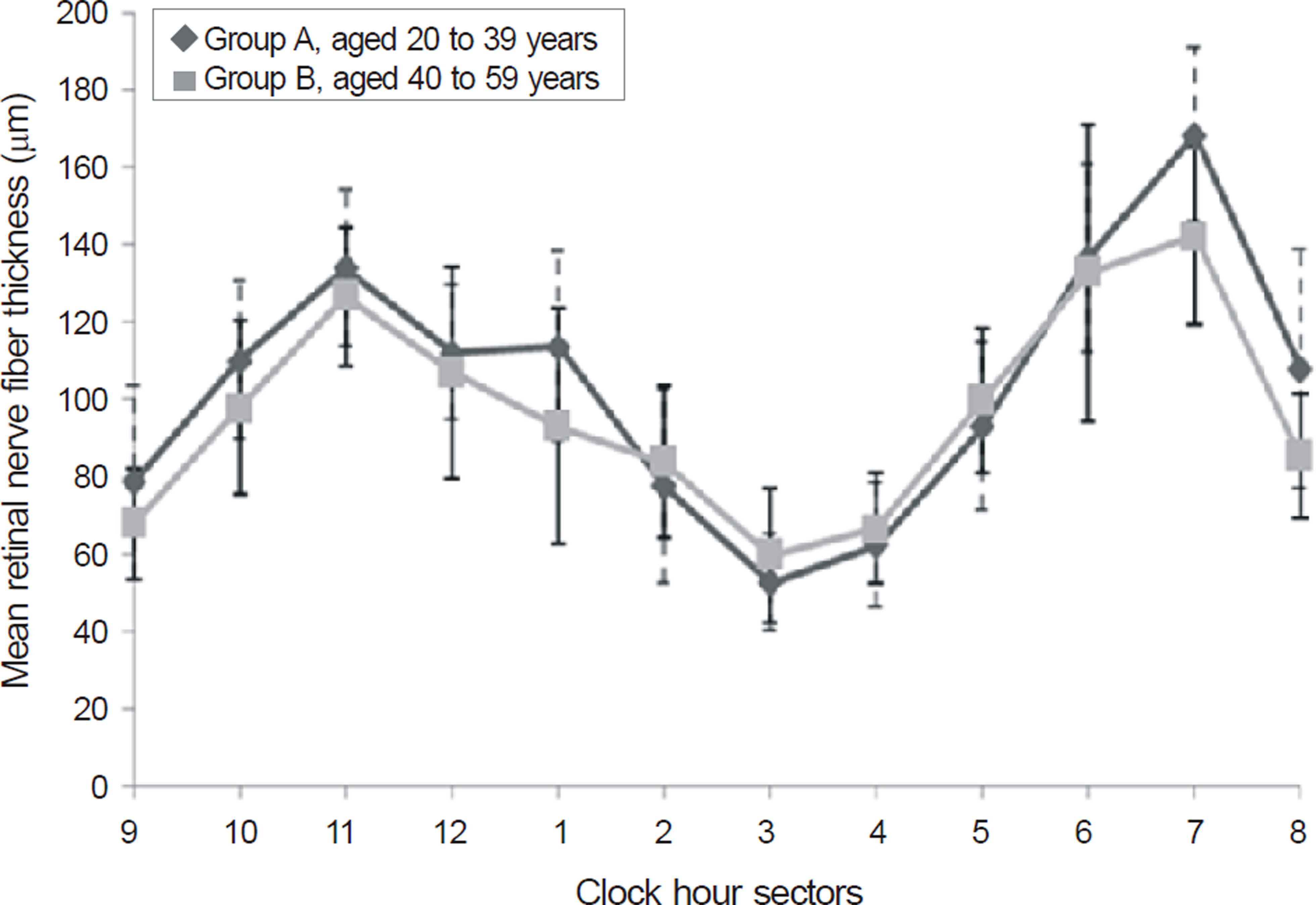
Figure 1. Comparison of retinal nerve fiber layer thickness profile between 2 groups. ◆(black diamond) = Group A, aged 20 to 39 years; ■(gray square) = Group B, aged 40 to 59 years. The standard deviation of each group was expressed with dotted (Group A) and straight lines (Group B). A sig-nificant difference of retinal nerve fiber layer thickness be-tween the young and old patients was revealed at 1 hour ( p = 0.020), 7 ( p = 0.001), and 8 ( p = 0.005) clock-hour sectors.
Reference
-
References
1. Katz J, Tielsch JM, Sommer A. Prevalence and risk factors for re-fractive errors in an adult inner city population. Invest Ophthalmol Vis Sci. 1997; 38:334–40.2. Wang Q, Klein BE, Klein R, Moss SE. Refractive status in the bea-ver dam eye study. Invest Ophthalmol Vis Sci. 1994; 35:4344–7.3. Lin LL, Shih YF, Hsiao CK, et al. Epidemiologic study of the prev-alence and severity of myopia among schoolchildren in taiwan in 2000. J Formos Med Assoc. 2001; 100:684–91.4. Wong TY, Foster PJ, Hee J, et al. Prevalence and risk factors for re-fractive errors in adult chinese in singapore. Invest Ophthalmol Vis Sci. 2000; 41:2486–94.5. Tay MT, Au Eong KG, Ng CY, Lim MK. Myopia and educational attainment in 421,116 young Singaporean males. Ann Acad Med Singapore. 1992; 21:785–91.6. Au Eong KG, Tay TH, Lim MK. Race, culture and myopia in 110,236 young Singaporean males. Singapore Med J. 1993; 34:29–32.7. Kang SH, Kim PS, Choi DG. Prevalence of myopia in 19-year-old Korean males: The relationship between the prevalence and educa-tion or urbanization. J Korean Ophthalmol Soc. 2004; 45:2082–7.8. Daubs JG, Crick RP. Effect of refractive error on the risk of ocular hypertension and open angle glaucoma. Trans Ophthalmol Soc U K. 1981; 101:121–6.9. Phelps CD. Effect of myopia on prognosis in treated primary open-angle glaucoma. Am J Ophthalmol. 1982; 93:622–8.
Article10. Podos SM, Becker B, Morton WR. High myopia and primary open-angle glaucoma. Am J Ophthalmol. 1966; 62:1038–43.
Article11. Perkins ES, Phelps CD. Open angle glaucoma, ocular hyper-tension, low-tension glaucoma, and refraction. Arch Ophthalmol. 1982; 100:1464–7.
Article12. Wilson MR, Hertzmark E, Walker AM, et al. A case-control study of risk factors in open angle glaucoma. Arch Ophthalmol. 1987; 105:1066–71.
Article13. Mitchell P, Hourihan F, Sandbach J, Wang JJ. The relationship be-tween glaucoma and myopia: The blue mountains eye study. Ophthalmology. 1999; 106:2010–5.14. Fong DS, Epstein DL, Allingham RR. Glaucoma and myopia: are they related? Int Ophthalmol Clin. 1990; 30:215–8.
Article15. Seddon JM, Schwartz B, Flowerdew G. Case-control study of ocu-lar hypertension. Arch Ophthalmol. 1983; 101:891–4.
Article16. Ko YC, Liu CJ, Chou JC, et al. Comparisons of risk factors and vis-ual field changes between juvenile-onset and late-onset primary open-angle glaucoma. Ophthalmologica. 2002; 216:27–32.
Article17. Tay E, Seah SK, Chan SP, et al. Optic disk ovality as an index of tilt and its relationship to myopia and perimetry. Am J Ophthalmol. 2005; 139:247–52.
Article18. Jonas JB, Gusek GC, Naumann GO. Optic disk morphometry in high myopia. Graefes Arch Clin Exp Ophthalmol. 1988; 226:587–90.
Article19. Schuman JS, Pedut-Kloizman T, Hertzmark E, et al. Reproducibility of nerve fiber layer thickness measurements using optical coher-ence tomography. Ophthalmology. 1996; 103:1889–98.
Article20. Blumenthal EZ, Williams JM, Weinreb RN, et al. Reproducibility of nerve fiber layer thickness measurements by use of optical co-herence tomography. Ophthalmology. 2000; 107:2278–82.21. Hoh ST, Greenfield DS, Mistlberger A, et al. Optical coherence to-mography and scanning laser polarimetry in normal, ocular hyper-tensive, and glaucomatous eyes. Am J Ophthalmol. 2000; 129:129–35.
Article22. Wollstein G, Schuman JS, Price LL, et al. Optical coherence to-mography (OCT) macular and peripapillary retinal nerve fiber lay-er measurements and automated visual fields. Am J Ophthalmol. 2004; 138:218–25.
Article23. Mohammad Salih PA. Evaluation of peripapillary retinal nerve fi-ber layer thickness in myopic eyes by spectral-domain optical co-herence tomography. J Glaucoma. 2012; 21:41–4.
Article24. Qiu KL, Zhang MZ, Leung CK, et al. Diagnostic classification of retinal nerve fiber layer measurement in myopic eyes: A compar-ison between time-domain and spectral-domain optical coherence tomography. Am J Ophthalmol. 2011; 152:646–53.e2.
Article25. Rauscher FM, Sekhon N, Feuer WJ, Budenz DL. Myopia affects retinal nerve fiber layer measurements as determined by optical co-herence tomography. J Glaucoma. 2009; 18:501–5.
Article26. Schweitzer KD, Ehmann D, Garcia R. Nerve fibre layer changes in highly myopic eyes by optical coherence tomography. Can J Ophthalmol. 2009; 44:e13–6.
Article27. Yoo YC, Lee CM, Park JH. Changes in peripapillary retinal nerve fi-ber layer distribution by axial length. Optom Vis Sci. 2012; 89:4–11.
Article28. Song TG, Yoo YC, Lee HB. Quantitative analysis of retinal nerve fiber layer thickness profile in myopic eyes. J Korean Ophthalmol Soc. 2009; 50:1840–46.
Article29. Leung CK, Mohamed S, Leung KS, et al. Retinal nerve fiber layer measurements in myopia: An optical coherence tomography study. Invest Ophthalmol Vis Sci. 2006; 47:5171–6.
Article30. Choi SW, Lee SJ. Thickness changes in the fovea and peripapillary retinal nerve fiber layer depend on the degree of myopia. Korean J Ophthalmol. 2006; 20:215–9.
Article31. Hougaard JL, Ostenfeld C, Heijl A, Bengtsson B. Modelling the normal retinal nerve fibre layer thickness as measured by stratus optical coherence tomography. Graefes Arch Clin Exp Ophthalmol. 2006; 244:1607–14.
Article32. Wollstein G, Schuman JS, Price LL, et al. Optical coherence to-mography (OCT) macular and peripapillary retinal nerve fiber lay-er measurements and automated visual fields. Am J Ophthalmol. 2004; 138:218–25.
Article33. Iliev ME, Meyenberg A, Garweg JG. Morphometric assessment of normal, suspect and glaucomatous optic discs with stratus OCT and HRT II. Eye (Lond). 2006; 20:1288–99.
Article34. Bayraktar S, Bayraktar Z, Yilmaz OF. Influence of scan radius cor-rection for ocular magnification and relationship between scan ra-dius with retinal nerve fiber layer thickness measured by optical coherence tomography. J Glaucoma. 2001; 10:163–9.
Article35. Kang SH, Hong SW, Im SK, et al. Effect of myopia on the thick-ness of the retinal nerve fiber layer measured by cirrus HD optical coherence tomography. Invest Ophthalmol Vis Sci. 2010; 51:4075–83.
Article36. Jonas JB, Schmidt AM, Muller-Bergh JA, et al. Human optic nerve fiber count and optic disc size. Invest Ophthalmol Vis Sci. 1992; 33:2012–8.37. Alamouti B, Funk J. Retinal thickness decreases with age: An OCT study. Br J Ophthalmol. 2003; 87:899–901.
Article38. Bowd C, Zangwill LM, Blumenthal EZ, et al. Imaging of the optic disc and retinal nerve fiber layer: The effects of age, optic disc area, refractive error, and gender. J Opt Soc Am A Opt Image Sci Vis. 2002; 19:197–207.
Article39. Hougaard JL, Kessel L, Sander B, et al. Evaluation of heredity as a determinant of retinal nerve fiber layer thickness as measured by optical coherence tomography. Invest Ophthalmol Vis Sci. 2003; 44:3011–6.
Article40. Kanamori A, Escano MF, Eno A, et al. Evaluation of the effect of aging on retinal nerve fiber layer thickness measured by optical co-herence tomography. Ophthalmologica. 2003; 217:273–8.
Article41. Yamada H, Yamakawa Y, Chiba M, Wakakura M. Evaluation of the effect of aging on retinal nerve fiber thickness of normal japanese measured by optical coherence tomography. Nihon Ganka Gakkai Zasshi. 2006; 110:165–70.42. Budenz DL, Anderson DR, Varma R, et al. Determinants of normal retinal nerve fiber layer thickness measured by stratus OCT. Ophthalmology. 2007; 114:1046–52.
Article
- Full Text Links
-
- Actions
-
Cited
- CITED
-
- Close
- Share
-
- Similar articles
-
- Reproducibility of Retinal Nerve Fiber Layer Thickness Evaluation by Nerve Fiber Analyzer
- Biometry of Retinal Nerve Fiber Layer Thickness by NFA
- Quantitative Analysis of Retinal Nerve Fiber Layer Thickness Profile in Myopic Eyes
- A Case of Retinal Herniation through Peripapillary Pit Resulting in Retinal Nerve Fiber Layer Defect
- Changes in Macular Retinal Layers and Peripapillary Nerve Fiber Layer Thickness after 577-nm Pattern Scanning Laser in Patients with Diabetic Retinopathy
