J Korean Ophthalmol Soc.
2013 Apr;54(4):675-679. 10.3341/jkos.2013.54.4.675.
A Case of Fungal Keratitis Scedosporium apiospermum
- Affiliations
-
- 1Department of Ophthalmology, Chonbuk National University Medical School, Jeonju, Korea. you2ic@paran.com
- 2Department of Labaratory Medicine, Chonbuk National University Medical School, Jeonju, Korea.
- KMID: 2216954
- DOI: http://doi.org/10.3341/jkos.2013.54.4.675
Abstract
- PURPOSE
To report a case of fungal keratitis caused by Scedosporium apiospermum.
CASE SUMMARY
A 70-year-old man visited our clinic with complaints of redness and decreased visual acuity in his right eye caused by a soil gotten into an eye while gardening 10 days ago. The patient had previously been treated in a local clinic but did not show significant clinical improvement. Bacterial and fungal staining, culture, and an antibiotic sensitivity test were performed from a corneal scrape. The cultures revealed growth of Scedosporium apiospermum. The patient was treated with topical moxifloxacin antibiotics, fluconazole, amphotericin B antifungal agents. However, the lesion was not improved, so antifungal therapy was switched to topical voriconazole. After two months of treatment, the infection was resolved with mild scarring.
CONCLUSIONS
Although it is a rare pathogen, Scedosporium apiospermum should be considered as a potential pathogen in patients presenting with corneal ulceration due to trauma from an object contaminated by soil, polluted water, or spoiled plant contact. And we suggest that topical application of voriconazole may be a good alternative treatment for patient with fungal keratitis in which no improvement despite a conventional antifungal agent, fluconazole.
MeSH Terms
Figure
-
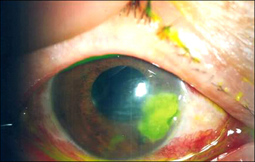
Figure 1. Corneal ulcer with deep stromal infiltration and surrounding edema were observed on initial presentation.
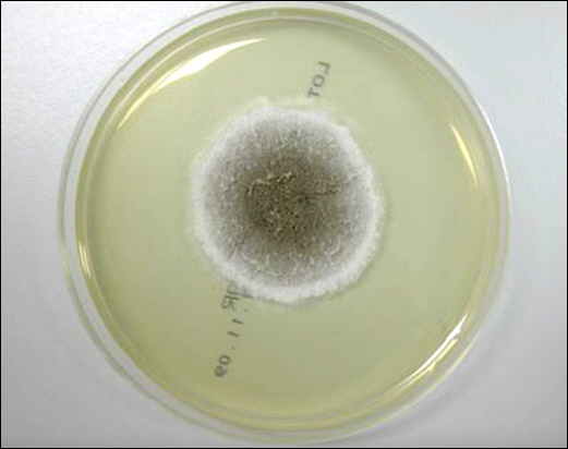
Figure 2. Dark graycolored cottony colony on Sabouraud dextrose agar after incubation.
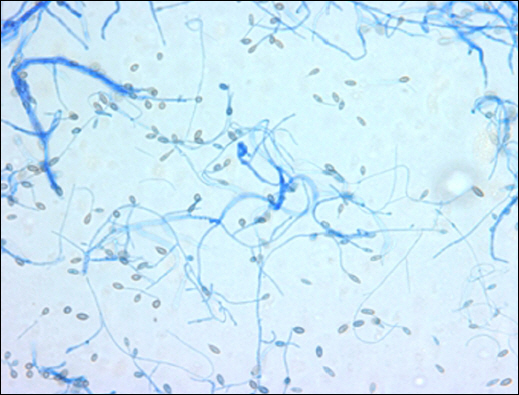
Figure 3. Microscopic morphology of Scedosporium apiospermum showing septate hyphae with simple long and short conidiophores (lactophenol cotton blue, ×;400).
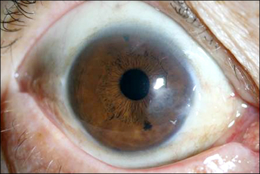
Figure 4. After 2 months, the corneal lesion regressed completely.
Reference
-
References
1. O'Day DM. Selection of appropriate antifungal therapy. Cornea. 1987; 6:238–45.2. Leber TH. Keratomycosis aspergillina als ursache von hypopyonkeratitis. Graefes Arch Ophthalmol. 1879; 25:285–301.
Article3. Jurkunas U, Behlau I, Colby K. Fungal keratitis: changing pathogens and risk factors. Cornea. 2009; 28:638–43.
Article4. Al-Badriyeh D, Leung L, Davies GE, et al. Successful salvage treatment of Scedosporium apiospermum keratitis with topical voriconazole after failure of natamycin. Ann Pharmacother. 2009; 43:1139–42.
Article5. Wu Z, Ying H, Yiu S, et al. Fungal keratitis caused by Scedosporium apiospermum: report of two cases and review of treatment. Cornea. 2002; 21:519–23.6. Thomas PA. Mycotic keratitisan underestimated mycosis. J Med Vet Mycol. 1994; 32:235–56.7. Cortez KJ, Roilides E, Quiroz-Telles F, et al. Infections caused by Scedosporium spp. Clin Microbiol Rev. 2008; 21:157–97.8. Yoon S, Kim S, Lee KA, Kim H. A case of Scedosporium apiospermum keratitis confirmed by a molecular genetic method. Korean J Lab Med. 2008; 28:307–11.
Article9. Ponchel C, Cassaing S, Linas MD, et al. [Fungal keratitis caused by Scedosporium apiospermum]. J Fr Ophthalmol. 2007; 30:933–7.10. Verweij PE, Brandt ME. Aspergillus, Fusarium, and other opportunistic moniliaceous fungi. Murray PR, Baron EJ, editors. Manual of Clinical Microbiology. 9th ed. Washington, DC: ASM Press;2007. p. 1821–2.11. Hahn YH, Hahn TW, Tchah H, et al. Epidemiology of infectious keratitis(II): a multi-center study. J Korean Ophthalmol Soc. 2001; 2:247–65.12. Ksiazek SM, Morris DA, Mandelbaum S, Rosenbaum PS. Fungal panophthalmitis secondary to Scedosporium apiospermum (Pseudallescheria boydii) keratitis. Am J Ophthalmol. 1994; 118:531–3.
Article13. Moshirfar M, Welling JD, Feiz V, et al. Infectious and non-infectious keratitis after laser in situ keratomileusis Occurrence, management, and visual outcomes. J Cataract Refract Surg. 2007; 3:474–83.14. Lalitha P, Prajna NV, Kabra A, et al. Risk factors for treatment outcome in fungal keratitis. Ophthalmology. 2006; 113:526–30.
Article15. Lee KH, Chae HJ, Yoon KC. Analysis of risk factors for treatment failure in fungal keratitis. J Korean Ophthalmol Soc. 2008; 49:37–42.
Article16. Sanati H, Belanger P, Fratti R, Ghannoum M. A new triazole, voriconazole (UK-109,496), blocks sterol biosynthesis in Candida albicans and Candida krusei. Antimicrob Agents Chemother. 1997; 1:2492–6.
Article17. Bunya VY, Hammersmith KM, Rapuano CJ, et al. Topical and oral voriconazole in the treatment of fungal keratitis. Am J Ophthalmol. 2007; 143:151–3.
Article18. Jhanji V, Yohendran J, Constantinou M, et al. Scedosporium scleritis or keratitis or both: case series. Eye Contact Lens. 2009; 5:312–5.
Article19. Srinivasan M. Fungal keratitis. Curr Opin Ophthalmol. 2004; 15:21–7.
Article
- Full Text Links
-
- Actions
-
Cited
- CITED
-
- Close
- Share
-
- Similar articles
-
- A Case of Scedosporium apiospermum Keratitis Confirmed by a Molecular Genetic Method
- A Case of Cutaneous Abscess Due to Scedosporium apiospermum
- A Case of Scedosporium Apiospermum Pneumonia in an Immunocompetent Patient
- Pulmonary Fungal Ball of Pseudallescheria boydiiIdentified by LSU rDNA D2 Region Sequencing
- A Case of Cutaneous Scedosporium apiospermum Infection
