The Effects of Pharmacologic Pupil Dilatation on Ocular, Corneal, and Internal Aberrations
- Affiliations
-
- 1Department of Ophthalmology, Korea University College of Medicine, Seoul, Korea. crisim@korea.ac.kr
- KMID: 2216938
- DOI: http://doi.org/10.3341/jkos.2013.54.4.581
Abstract
- PURPOSE
The present study investigates the effects of pharmacologic pupil dilatation on ocular, corneal and internal aberrations.
METHODS
Sixty-two right eyes of 62 healthy participants were included in the present study. Ocular, corneal and internal aberrations were measured with a KR-1W wavefront aberrometer (Topcon Corp., Tokyo, Japan) before mydriasis in mesopic conditions. After pupil dilatation with a mydriatic drug (phenylephrine chloride 0.5% + tropicamide 0.5%) (Mydrin-P, Santen, Osaka, Japan), the measurements were repeated. The wavefront data of 4-mm and 6-mm diameter zones were analyzed. The changes of aberrations before and after mydriasis were evaluated by paired t-test.
RESULTS
The values of ocular, corneal and internal spherical aberrations before and after mydriasis on the 4-mm diameter pupil zone were not statistically significantly different. On the 6-mm diameter zone, the ocular and internal spherical aberrations were statistically significantly different (p = 0.025, p = 0.002, respectively, paired t-test). However, the corneal aberrations did not show significant changes. The internal aberrations average before mydriasis was -0.043 (+/-0.21) microm and was shifted in a negative direction to -0.093 (+/-0.17) microm after mydriasis. The ocular aberrations average also changed toward negative after mydriasis. The high-order aberrations and astigmatism did not change significantly.
CONCLUSIONS
The ocular and internal spherical aberrations changed toward negative with mydriasis in the participants' eyes suggesting the change of the ocular spherical aberration to be attributed to internal changes.
Keyword
Figure
-

Figure 1. Comparison of spherical aberrations (SA) before and after mydriasis (in micrometer). The ocular and internal spherical aberrations were statistically significantly different on 6-mm diameter zone (p = 0.025, p = 0.002, respectively, paired t-test). Before = before mydriasis, at mesopic conditions, 8 lux; After = after pharmacologically induced mydriasis.
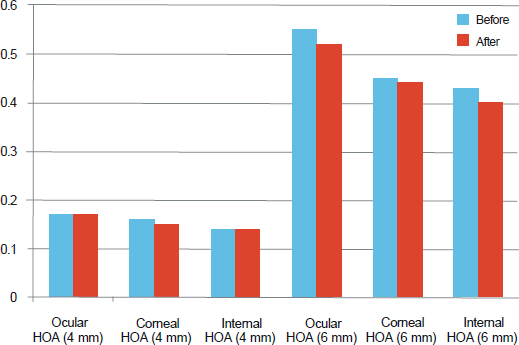
Figure 2. Comparison of high order aberrations (HOA) before and after mydriasis (in average root mean square; RMS). The high order aberrations did not change significantly after mydriasis. Before = before mydriasis, at mesopic conditions, 8 lux; After = after pharmacologically induced mydriasis.
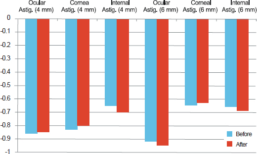
Figure 3. Comparison of astigmatism (Astig.) before and after mydriasis (in diopter). The astigmatism did not change significantly after mydriasis. Before = before mydriasis, at mesopic conditions, 8 lux; After = after pharmacologically induced mydriasis.
Cited by 1 articles
-
Analysis of Internal Optical Aberrations in Eyes with Different Types of Cataract
Ji Yun Han, Young Sub Eom, Jay Won Rhim, Su Yeon Kang, Hyo Myung Kim, Jong Suk Song
J Korean Ophthalmol Soc. 2015;56(4):532-540. doi: 10.3341/jkos.2015.56.4.532.
Reference
-
References
1. Cashell GT. A short history of spectacles. Proc R Soc Med. 1971; 64:1063–4.
Article2. Born M, Wolf E. Principles of optics, 7th ed. Cambridge: Cambridge University Press;1999. p. 523–5.3. Molebny VV, Pallikaris IG, Naoumidis LP, et al. Retina ray-tracing technique for eye-refraction mapping. Proc SPIE. 1997; 2971:175–83.4. Molebny VV, Panagopoulou SI, Molebny SV, et al. Principles of ray tracing aberrometry. J Refract Surg. 2000; 16:S572–5.
Article5. MacRae S, Fujieda M. Slit skiascopic-guided ablation using the Nidek laser. J Refract Surg. 2000; 16:S576–80.
Article6. Mrochen M, Kaemmerer M, Mierdel P, et al. Principles of Tscherning aberrometry. J Refract Surg. 2000; 16:S570–1.
Article7. Moreno-Barriuso E, Navarro R. Laser Ray Tracing versus Hartmann-Shack sensor for measuring optical aberrations in the human eye. J Opt Soc Am A Opt Image Sci Vis. 2000; 17:974–85.
Article8. Thibos LN. Principles of Hartmann-Shack aberrometry. J Refract Surg. 2000; 16:S563–5.
Article9. Liang J, Grimm B, Goelz S, Bille JF. Objective measurement of wave aberrations of the human eye with the use of a Hartmann-Shack wave-front sensor. J Opt Soc Am A Opt Image Sci Vis. 1994; 11:1949–57.
Article10. Ortiz D, Alió JL, Bernabéu G, Pongo V. Optical performance of monofocal and multifocal intraocular lenses in the human eye. J Cataract Refract Surg. 2008; 34:755–62.
Article11. Giessler S, Hammer T, Duncker GI. [Aberrometry due dilated pupilsWhich mydriatic should be used?]. Klin Monbl Augenheilkd. 2002; 219:655–9.12. Carkeet A, Velaedan S, Tan YK, et al. Higher order ocular aberrations after cycloplegic and non-cycloplegic pupil dilation. J Refract Surg. 2003; 19:316–22.
Article13. Taneri S, Oehler S, Azar DT. Influence of mydriatic eye drops on wavefront sensing with the Zywave aberrometer. J Refract Surg. 2011; 27:678–85.
Article14. Ahn SM, Seok SS, Park CY. Considering spherical aberration in choosing the wavefront map for laser vision correction. J Korean Ophthalmol Soc. 2011; 52:147–56.
Article15. Piñero DP, Juan JT, Alió JL. Intrasubject repeatability of internal aberrometry obtained with a new integrated aberrometer. J Refract Surg. 2011; 27:509–17.
Article16. Brown N. The change in shape and internal form of the lens of the eye on accommodation. Exp Eye Res. 1973; 15:441–59.
Article17. Dubbelman M, Van der Heijde GL, Weeber HA. Change in shape of the aging human crystalline lens with accommodation. Vision Res. 2005; 45:117–32.
Article18. Dubbelman M, Van der Heijde GL, Weeber HA, Vrensen GF. Changes in the internal structure of the human crystalline lens with age and accommodation. Vision Res. 2003; 43:2363–75.
Article
- Full Text Links
-
- Actions
-
Cited
- CITED
-
- Close
- Share
-
- Similar articles
-
- Comparison between Anterior Corneal Aberration and Ocular Aberration in Laser Refractive Surgery
- Wavefront Analysis of Successful Treatment of Monocular Triplopia After Cataract Extraction: Report of 2 Cases
- The Changes of Corneal and Ocular High-Order Aberrations before and after Playing Computer Games
- Comparison of Ocular Aberration and Clinical Outcome between Different Aspheric Intraocular Lenses in Both Eyes
- Comparison of Internal and Total Optical Aberrations for 2 Aberrometers: iTrace and OPD Scan
