Intravitreal Bevacizumab Injection for Serous Retinal Detachment Associated with Leber's Idiopathic Stellate Neuroretinitis
- Affiliations
-
- 1Department of Ophthalmology, Gangnam Severance Hospital, Yonsei University College of Medicine, Seoul, Korea. minkim76@yuhs.ac
- 2Department of Ophthalmology, Severance Hospital, Yonsei University College of Medicine, Seoul, Korea.
- KMID: 2216880
- DOI: http://doi.org/10.3341/jkos.2014.55.10.1562
Abstract
- PURPOSE
To report the effectiveness of intravitreal bevacizumab treatment for serous retinal detachment associated with Leber's idiopathic stellate neuroretinitis.
CASE SUMMARY
A 56-year-old male visited our clinic complaining of visual disturbance for three days in his right eye. His best corrected visual acuity was 0.5. Relative afferent pupillary defect and pain when moving eyes were noted in his right eye as well as inflammatory cells in the vitreous cavity. On funduscopic examination, disc swelling with hemorrhage and stellate-shaped hard exudates were noted at the perifovea. Ishihara color vision test showed anomalous trichromacy in his right eye. Hyperfluorescence around the disc was observed on fundus fluorescein angiography. Optical coherence tomography showed disc swelling with serous retinal detachment at the fovea. Inferior altitudinal scotoma was noted on visual field examination. The patient underwent intravitreal bevacizumab injection and topical steroid medication. After six days, the patient's symptoms and disc swelling improved, and decreased subretinal fluid was observed. After six weeks, his best corrected visual acuity was 1.0. Nine weeks later, visual field examination showed nonspecific scotoma.
CONCLUSIONS
Intravitreal bevacizumab injection is useful for treating serous retinal detachment associated with Leber's idiopathic stellate neuroretinitis.
Keyword
MeSH Terms
Figure
-
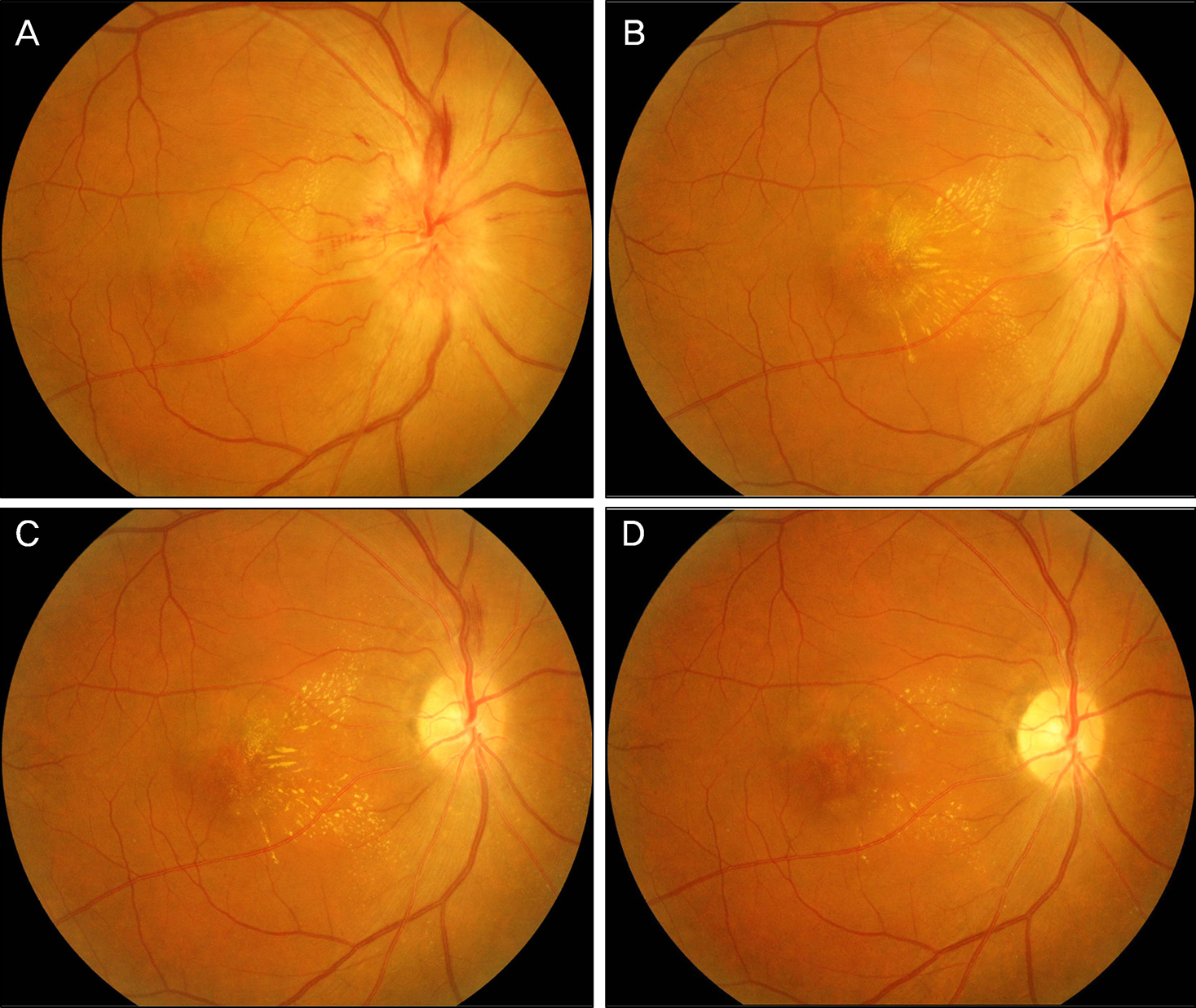
Figure 1. Consecutive fundus color photographs of the patient. At the initial visit, disc swelling with blurring of the disc margin, disc hemorrhage, and stellate-shaped hard exudates at the perifovea are noted (A). Six days after the first injection of bevacizumab, improvement of disc swelling but slight increased exudates at the fovea was noted (B). Six weeks after injection of bevacizumab (C). Note the resolution of disc swelling and improvement of stellate-shaped hard exudates at the perifovea (C). Fifteen weeks after the first visit, disc swelling with hemorrhage is completely regressed, and hard exudates remaining at the perifovea are markedly decreased compared to the fundus color photograph taken at 6 days after the first injection of bevacizumab (D).
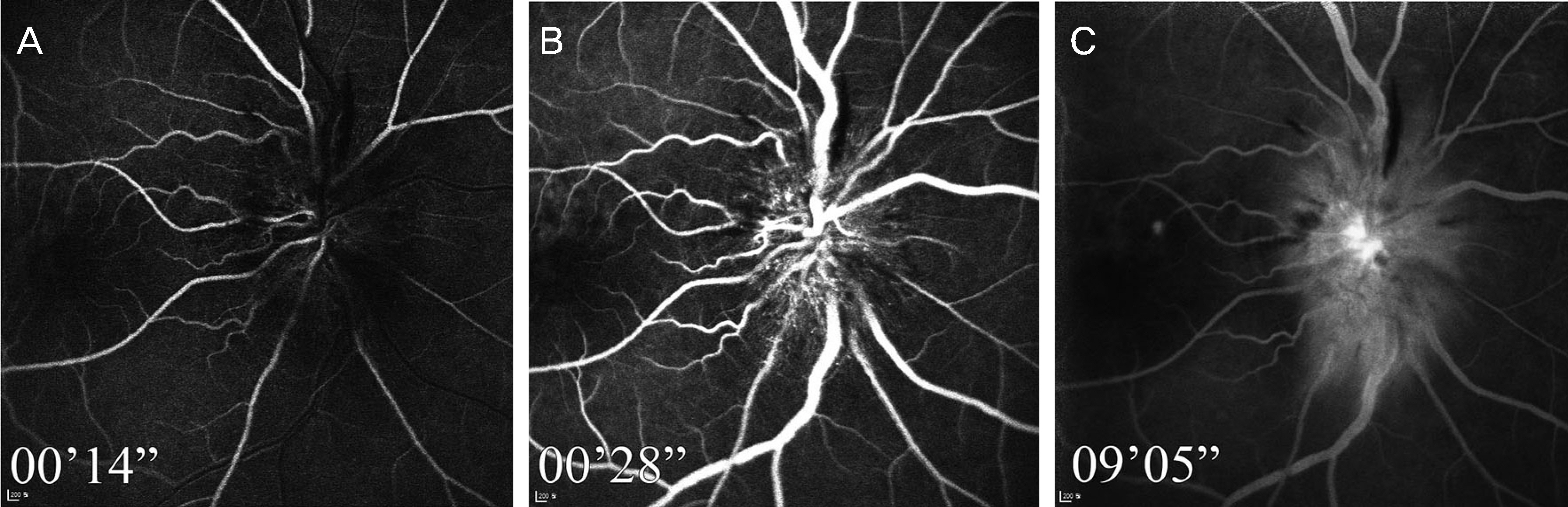
Figure 2. Fundus fluorescein angiography at the initial visit. Early hyperfluorescence at the optic disc continues until the late phase. Fundus fluorescein angiography at the initial visit (A, B, C). Early hyperfluorescence at the optic disc (A) continues until the late phase (B, C).
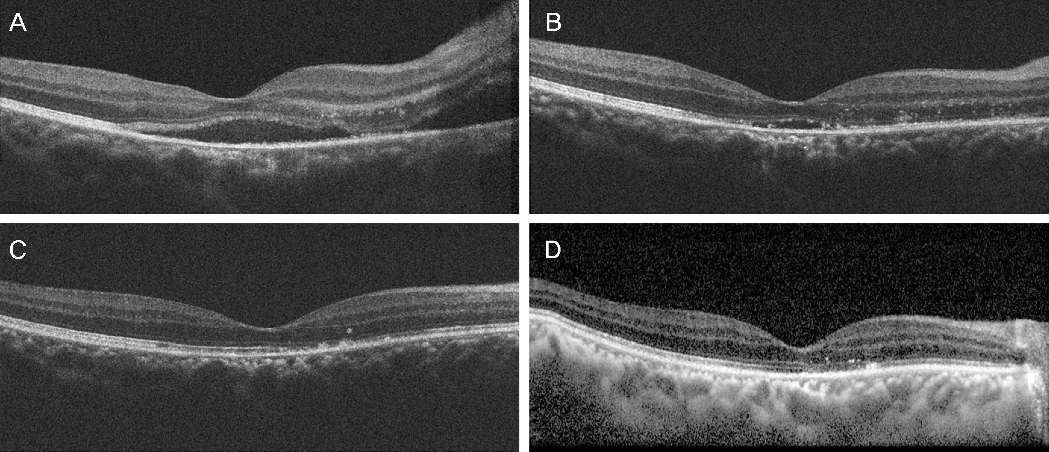
Figure 3. Consecutive optical coherence tomography (OCT) images of the patient. Serous retinal detachment at the fovea and around the disc with subretinal fluid is noted (A). Six days after injection of bevacizumab, improvement of serous retinal detachment with decreased amount of subretinal fluid is noted (B). Six weeks after injection of bevacizumab, note the complete resolution of subretinal fluid and disruption of inner segment/outer segment (IS/OS) junction line at the nasal perifovea (C). Fifteen weeks after the first visit, disrupted IS/OS junction line at the nasal perifovea shows more improvement compared to the OCT image taken at 6 weeks after injection of bevacizumab (D).

Figure 4. Automated visual field at the initial visit (A) and after 9 weeks (B). Although the reliability of the examination at 9 weeks is lower inferior altitudinal scotoma had disappeared at 9 weeks, compared to the initial visual field. POS = positive; NEG = negative; ASB = apostilbs; RX = prescription; DS = diopter sphere; DC = diopter cylinder; X = axis; GHT = glaucoma hemifield test; VFI = visual field index; MD = mean deviation; PSD = pattern standard deviation.
Cited by 1 articles
-
Clinical Manifestation and Outcomes of Neuroretinitis in Korea
Su Gyeong Jang, Kang Yeun Pak, Han Jo Kwon, Seung Min Lee, Sung Who Park, Ik Soo Byon, Ji Eun Lee
J Korean Ophthalmol Soc. 2017;58(2):156-164. doi: 10.3341/jkos.2017.58.2.156.
Reference
-
References
1. Purvin V, Sundaram S, Kawasaki A. Neuroretinitis: review of the literature and new observations. J Neuroophthalmol. 2011; 31:58–68.
Article2. Suhler EB, Lauer AK, Rosenbaum JT. Prevalence of serologic evidence of cat scratch disease in patients with neuroretinitis. Ophthalmology. 2000; 107:871–6.3. T L. Die pseudonephritischen Netzhauterkrnakungen, die Retinitis stellata: Die Purtschersche Netzhauteffektion nach schwerer Schadelverletzung. Graefe AC, Saemisch T, editors. Graefe-Saemisch Handbuch der Gesamten Augerheikunde. 2nd ed.Leipzig: Englemann;1916. 78.4. Avery RL, Pieramici DJ, Rabena MD, et al. Intravitreal bevacizumab (Avastin) for neovascular age-related macular degeneration. Ophthalmology. 2006; 113:363–72.e5.
Article5. Avery RL, Pearlman J, Pieramici DJ, et al. Intravitreal bevacizumab (Avastin) in the treatment of proliferative diabetic retinopathy. Ophthalmology. 2006; 113:1695.e1–15.
Article6. Akesbi J, Brousseaud FX, Adam R, et al. Intravitreal bevacizumab (Avastin) in idiopathic retinitis, vasculitis, aneurysms and neuroretinitis. Acta Ophthalmol. 2010; 88:e40–1.
Article7. Sawhney GK, Payne JF, Ray R, et al. Combination anti-VEGF and corticosteroid therapy for idiopathic retinal vasculitis, aneurysms, and neuroretinitis syndrome. Ophthalmic Surg Lasers Imaging Retina. 2013; 44:599–602.
Article8. Jung JJ, Baek SH, Kim US. Analysis of the causes of optic disc swelling. Korean J Ophthalmol. 2011; 25:33–6.
Article9. Han DH, Sohn HJ, Lee DY, Nam DH. Leber's idiopathic stellate neuroretinitis with peripapillary serous retinal detachment. J Korean Ophthalmol Soc. 2011; 52:1109–13.
Article10. Empeslidis T, Banerjee S, Vardarinos A, Konstas AG. Dexamethasone intravitreal implant for idiopathic retinal vasculitis, aneurysms, and neuroretinitis. Eur J Ophthalmol. 2013; 23:757–60.
Article11. Erdurman FC, Durukan AH, Mumcuoğlu T, Hürmeriç V. Intravitreal bevacizumab treatment of macular edema due to optic disc vasculitis. Ocul Immunol Inflamm. 2009; 17:56–8.
Article12. Finger PT. Anti-VEGF bevacizumab (Avastin) for radiation optic neuropathy. Am J Ophthalmol. 2007; 143:335–8.
Article13. Bennett JL, Thomas S, Olson JL, Mandava N. Treatment of nonarteritic anterior ischemic optic neuropathy with intravitreal bevacizumab. J Neuroophthalmol. 2007; 27:238–40.
Article14. Al-Dhibi H, Khan AO. Response of diabetic papillopathy to intravitreal bevacizumab. Middle East Afr J Ophthalmol. 2011; 18:243–5.
Article15. Cakir M, Cekiç O, Bozkurt E, et al. Combined intravitreal bevacizumab and triamcinolone acetonide injection for idiopathic neuroretinitis. Ocul Immunol Inflamm. 2009; 17:221–3.16. Rappoport D, Morzaev D, Weiss S, et al. Effect of intravitreal injection of bevacizumab on optic nerve head leakage and retinal ganglion cell survival in a mouse model of optic nerve crush. Invest Ophthalmol Vis Sci. 2013; 54:8160–71.
Article
- Full Text Links
-
- Actions
-
Cited
- CITED
-
- Close
- Share
-
- Similar articles
-
- Leber's Idiopathic Stellate Neuroretinitis with Peripapillary Serous Retinal Detachment
- The Short-term Effect of Intravitreal Bevacizumab for Treatment of Central Serous Chorioretinopathy
- Serous Retinal Detachment Following Combined Photodynamic Therapy and Intravitreal Bevacizumab Injection
- A Case of Central Serous Chorioretinopathy Associated With Retinal Detachment Improved by Intravitreal Bevacizumab Injection
- A Case of Intravitreal Bevacizumab Injection for the Treatment of Choroidal Neovascularization in Morning Glory Syndrome
