J Korean Ophthalmol Soc.
2013 Jan;54(1):123-130. 10.3341/jkos.2013.54.1.123.
Visual Field Relocation and Clinical Effect of Fresnel Prism in Patients with Homonymous Hemianopsia
- Affiliations
-
- 1Department of Ophthalmology, Chung-Ang University College of Medicine, Seoul, Korea. njmoon@chol.com
- KMID: 2216460
- DOI: http://doi.org/10.3341/jkos.2013.54.1.123
Abstract
- PURPOSE
To report the result of Fresnel prism application and adaptation for visual field relocation and functional vision improvement in homonymous hemianopsia patients.
METHODS
Fifteen homonymous hemianopsia patients were prescribed Fresnel prism. To expand the visual field, Fresnel prism was placed base-out toward the defective field and patients were given an adaptation period of 1 month. The effects of the prism on field expansion was evaluated using Goldmann perimetry. In addition, the NEI-VFQ25 questionnaire was utilized asking patients regarding their subjective functional vision and satisfaction in daily life before and after using the Fresnel prism.
RESULTS
After 1 month of Fresnel prism prescription, 53% of patients showed objective visual field expansion to the defective field of 12.5 degrees on average. Monocular or macular splitting hemianopsia patients showed more visual field expansion than binocular macular sparing hemianopsia patients. The NEI-VFQ25 score increased significantly and abnormal head position decreased or disappeared after 1 month of using the prism. However, 47% of patients failed to adapt to the prism.
CONCLUSIONS
Using Fresnel prism in homonymous hemianopsia patients effectively expands the visual field, corrects abnormal head position, and improves functional vision. However, to improve the success rate, for certain patients the proper choice of prism application method, prism diopters, and constant management are necessary.
Keyword
Figure
-
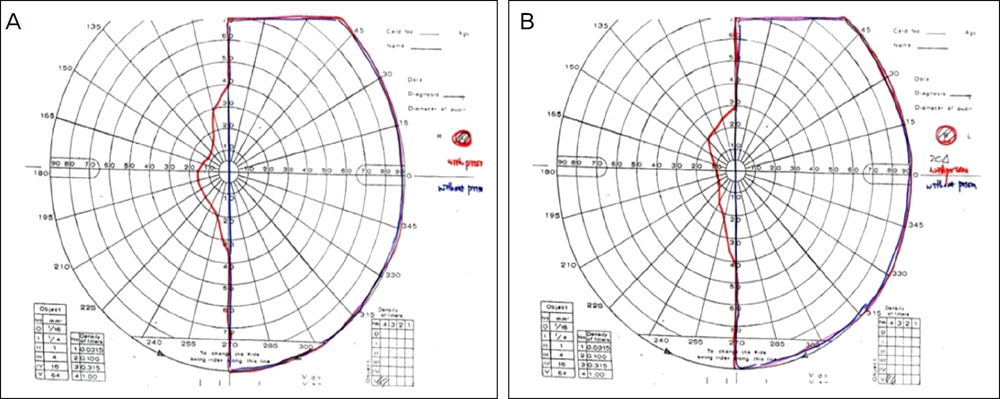
Figure 1. Visual fields have been expanded from the seeing side into the defected side after using prisms on a 27 years old male patient with left side homonymous hemianopsia. Prisms were prescribed as right side 20 prism diopters base-in, and left side 20 prism diopters base-out (Blue line: Before using prism, Red line: After using prism, (A) Visual field of the right eye, (B) Visual field of the left eye).
Reference
-
References
1. Gilhotra JS, Mitchell P, Healey PR, et al. Homonymous visual field defects and stroke in an older population. Stroke. 2002; 33:2417–20.
Article2. Zhang X, Kedar S, Lynn MJ, et al. Homonymous hemianopias – clinical-anatomic correlations in 904 cases. Neurology. 2006; 66:906–10.3. Rossi PW, Kheyfets S, Reding MJ. Fresnel prisms improve visual perception in stroke patients with homonymous hemianopia or unilateral visual neglect. Neurology. 1990; 40:1597–9.
Article4. Riss-Jayle M, Giorgi R, Barthes A. [Setting the preferential retinal locus. Part 1. Analysis of the rehabilitation results as a function of positioning]. J Fr Ophtalmol. 2008; 31:249–55.5. Prsa M, Galiana HL. Visual-Vestibular interaction hypothesis for the control of orienting gaze shifts by brain stem omnipause neurons. J Neurophysiol. 2007; 97:1149–62.
Article6. Kasten E, Wüst S, Behrens-Baumann W, Sabel BA. Computerbased training for the treatment of partial blindness. Nat Med. 1998; 4:1083–7.
Article7. Kerkhoff G, Münßinger U, Haaf E, et al. Rehabilitation of abdominal scotomata in patients with postgeniculate damage of the visual system: saccadic compensation training. Restor Neurol Neurosci. 1992; 4:245–54.8. Kerkhoff G, Münßinger U, Meier EK. Neurovisual rehabilitation in cerebral blindness. Arch Neurol. 1994; 51:474–81.
Article9. Nelles G, Esser J, Eckstein A, et al. Compensatory visual field training for patients with hemianopia after stroke. Neurosci Lett. 2001; 306:189–92.
Article10. Pambakian AL, Mannan SK, Hodgson TL, Kennard C. Saccadic visual search training: a treatment for patients with homonymous hemianopia. J Neurol Neurosurg Psychiatry. 2004; 75:1443–8.
Article11. Al-Karmi R, Markowitz SN. Image relocation with prisms in abdominals with age-related macular degeneration. Can J Ophthalmol. 2006; 41:313–8.12. Quah SA, Kaye SB. Binocular visual field changes after surgery in esotropic amblyopia. Invest Ophthalmol Vis Sci. 2004; 45:1817–22.
Article13. Peli E. Vision multiplexing: an engineering approach to vision abdominal device development. Optom Vis Sci. 2001; 78:304–15.14. Heo JW, Yoon HS, Shin JP, et al. A validation and reliability study of the Korean version of national eye institute visual function questionnaire 25. J Korean Ophthalmol Soc. 2010; 51:1354–67.
Article15. Giorgi RG, Woods RL, Peli E. Clinical and laboratory evaluation of peripheral prism glasses for hemianopia. Optom Vis Sci. 2009; 86:492–502.
Article16. Bowers AR, Keeney K, Peli E. Community-based trial of a abdominal prism visual field expansion device for hemianopia. Arch Ophthalmol. 2008; 126:657–64.17. Gassel MM, Williams D. Visual function in patients with abdominal hemianopial II Oculomotor mechanisms. Brain. 1963; 86:1–36.18. Martin T, Riley ME, Kelly KN, et al. Visually-guided behavior of homonymous hemianopes in a naturalistic task. Vision Res. 2007; 47:3434–46.
Article
