J Korean Ophthalmol Soc.
2012 Sep;53(9):1357-1363. 10.3341/jkos.2012.53.9.1357.
A Case of Hydroxychloroquine Retinopathy
- Affiliations
-
- 1Myung-Gok Eye Research Institute, Department of Ophthalmology, Kim's Eye Hospital, Konyang University, Seoul, Korea. yousujin@kimeye.com
- 2Department of Ophthalmology, Konyang University College of Medicine, Daejeon, Korea.
- KMID: 2216093
- DOI: http://doi.org/10.3341/jkos.2012.53.9.1357
Abstract
- PURPOSE
Hydroxychloroquine has been used as the antimalarial agent and drug of the treatment for autoimmune disease such as rheumatoid arthritis. Hydroxychloroquine retinopathy can cause serious visual disturbance although the incidence is low. This report is to describe a case of Hydroxycholoroquine retinopathy on 73 year old female.
CASE SUMMARY
A 73 year old female patient presented our clinic with complaints of visual disturbance for several months. She had taking 400 mg/day (8.8 mg/kg of lean body weight/day) of hydroxychloroquine for 2 years. The best corrected visual acuity was 20/30 in both eyes. Bull's eye maculopathy was observed on her fundus examination and Humphrey Automated Visual Field 24-2 showed central scotoma in both eyes. Parafoveal thinning of photoreceptor layers, loss of the inner and outer segment junction and external limiting membrane was observed on spectral domain Optical Coherence Tomography. Window defect was visible at the parafoveal area on fluorescein angiography. Electroretinogram revealed subtle dysfunction of cone cell and multifocal ERG trace array showed decreased amplitudes at the parafoveal area. Electrooculogram showed decreased Arden ratio.
CONCLUSIONS
We strongly advise that all patients taking Hydorxycholoroquine therapy have a regular examination to find hydroxychloroquine retinopathy in early stage especially in the patients having high risk factors.
MeSH Terms
Figure
-

Figure 1 Fundus photograph shows diffuse RPE atrophy of macula with foveal sparing in both eyes. (A) Right eye. (B) Left eye.

Figure 2 Humphrey Automated Visual Field 24-2 shows central scotoma in both eyes.

Figure 3 Spectral domain-OCT shows parafoveal thinning of photoreceptor layers and loss of the inner and outer segment junction line (white arrows).
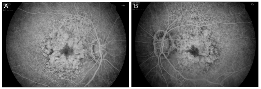
Figure 4 Fluorescein Angiography shows window defect at parafoveal area (Bull's eye maculopathy). (A) Right eye. (B) Left eye.
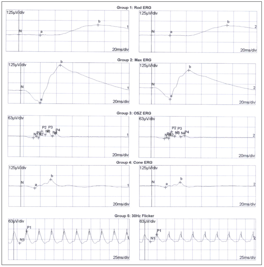
Figure 5 Electroretinogram reveals subtle dysfunction of cone cell.
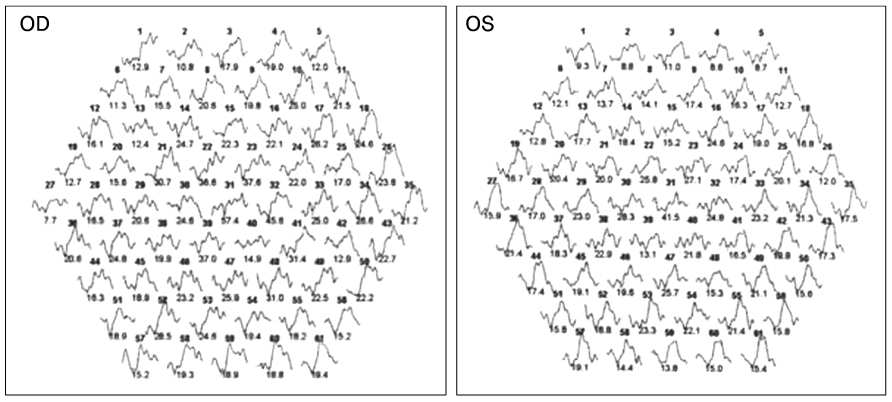
Figure 6 Multifocal ERG trace array shows decreased amplitudes in parafoveal area.

Figure 7 Electrooculogram shows decreased Arden ratio.
Reference
-
1. Hobbs HE, Sorsby A, Freedman A. Retinopathy following chloroquine therapy. Lancet. 1959. 2:478–480.2. Shearer RV, Dubois EL. Ocular changes induced by long-term hydroxychloroquine (plaquenil) therapy. Am J Ophthalmol. 1967. 64:245–252.3. Johnson MW, Vine AK. Hydroxychloroquine therapy in massive total doses without retinal toxicity. Am J Ophthalmol. 1987. 104:139–144.4. Mills PV, Beck M, Power BJ. Assessment of the retinal toxicity of hydroxychloroquine. Trans Ophthalmol Soc U K. 1981. 101:109–113.5. Klinger G, Morad Y, Westall CA, et al. Ocular toxicity and antenatal exposure to chloroquine or hydroxychloroquine for rheumatic diseases. Lancet. 2001. 358:813–814.6. Shroyer NF, Lewis RA, Lupski JR. Analysis of the ABCR (ABCA4) gene in 4-aminoquinoline retinopathy: is retinal toxicity by chloroquine and hydroxychloroquine related to Stargardt disease? Am J Ophthalmol. 2001. 131:761–766.7. Maturi RK, Folk JC, Nichols B, et al. Hydroxychloroquine retinopathy. Arch Ophthalmol. 1999. 117:1262–1263.8. Maturi RK, Yu M, Weleber RG. Multifocal electroretinographic evaluation of long-term hydroxychloroquine users. Arch Ophthalmol. 2004. 122:973–981.9. Marmor MF, Kellner U, Lai TY, et al. Revised recommendations on screening for chloroquine and hydroxychloroquine retinopathy. Ophthalmology. 2011. 118:415–422.10. Pinckers A, Cruysberg JR, aan de Kerk AL. Main types of bull's eye maculopathy. Functional classification. Doc Ophthalmol. 1984. 58:257–267.11. Stepien KE, Han DP, Schell J, et al. Spectral-domain optical coherence tomography and adaptive optics may detect hydroxychloroquine retinal toxicity before symptomatic vision loss. Trans Am Ophthalmol Soc. 2009. 107:28–33.12. Bergholz R, Schroeter J, Rüther K. Evaluation of risk factors for retinal damage due to chloroquine and hydroxychloroquine. Br J Ophthalmol. 2010. 94:1637–1642.
- Full Text Links
-
- Actions
-
Cited
- CITED
-
- Close
- Share
-
- Similar articles
-
- Hydroxychloroquine Retinopathy
- Prevalence and Risk Factors of Hydroxychloroquine Retinopathy in Rheumatic Patients with Dry Eye Symptoms
- Frequency and Clinical Characteristics of Hydroxychloroquine Retinopathy in Korean Patients with Rheumatologic Diseases
- A Case of Recurrent Annular Erythema of Sjogren's Syndrome Treated with Hydroxychloroquine
- Hydroxychloroquine Retinopathy Update
