A Case of Carcinoma Ex Pleomorphic Adenoma at 1st Operation
- Affiliations
-
- 1Department of Ophthalmology and Visual Science, The Catholic University of Korea College of Medicine, Seoul, Korea. yswoph@hanmail.net
- KMID: 2215712
- DOI: http://doi.org/10.3341/jkos.2015.56.4.598
Abstract
- PURPOSE
To report a case of carcinoma ex pleomorphic adenoma observed during the patient's first operation.
CASE SUMMARY
A 63-year-old female presented with proptosis and ptosis that was aggravated 1 year prior. On preoperative CT image, a 32 x 20 x 21 mm-sized well demarcated mass (suspected as pleomorphic adenoma) was observed and was removed entirely by anterolateral orbitotomy. The excised mass surface was uneven but the capsule appeared intact on gross examination. Hard, yellow-colored and soft, dark-colored materials were found concurrently on cross section. The histological examination showed malignant cells as part of the soft material and was diagnosed as carcinoma ex pleomorphic adenoma.
CONCLUSIONS
We report a case of carcinoma ex pleomorphic adenoma of the lacrimal gland that presented with malignant change during the patient's first operation. Supposedly, during the process of mass growth, minimal rupture occurred causing malignant transformation. Clinically, although a mass is believed benign based on imaging, the possibility of malignant transformation of a tumor increasing rapidly or enlargement causing development of rapid proptosis should be considered.
Keyword
MeSH Terms
Figure
-
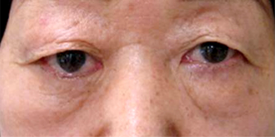
Figure 1. The patient shows ptosis of right eye at primary position in preoperative frontal view.

Figure 2. On CT image, 32 × 20 × 21 mm sized well dermacated mass is seen (A: coronal view; B: axial view).

Figure 3. On cross sectional photo of excised mass (A), the pathologic finding of upper- nodular, translucent lesion has benign characteristics (white arrow), and lower- tan-yellow, firm mass with ill-defined lesion has malignant characterisctics (black arrow). The malignant component is distinguished by its collagenous stromal hyalinization. On microscopic examination (B), morphologic diver-sity with both epithelioid and connective tissue components is seen, which is consistent with pleomorphic adenoma (white arrow). This component is partially replaced by the malignant component (black arrow) – duct formation with nucleus containing chromatin condensation and prominent nucleoli is seen on high power, which is consistent with adenocarcinoma.

Figure 4. After operation, ptosis is improved in frontal view.
Reference
-
References
1. Shields JA, Shields CL, Scartozzi R. Survey of 1264 patients with orbital tumors and simulating lesions: The 2002 Montgomery Lecture, part 1. Ophthalmology. 2004; 111:997–1008.2. Jung BJ, Cho YK, La TY. A giant pleomorphic adenoma of lacrimal gland involving the palpebral lobe causing severe mechan-ical ptosis. J Korean Ophthalmol Soc. 2011; 52:241–5.
Article3. Shields JA, Shields CL, Eagle RC, Rizzo J. Pleomorphic adenoma (‘benign mixed tumor’) of the lacrimal gland. Arch Ophthalmol. 1987; 105:560–1.
Article4. von Holstein SL, Fehr A, Persson M, et al. Lacrimal gland pleomorphic adenoma and carcinoma ex pleomorphic adenoma: genomic profiles, gene fusions, and clinical characteristics. Ophthalmology. 2014; 121:1125–33.5. von Holstein SL. Tumours of the lacrimal gland. Epidemiological, clinical and genetic characteristics. Acta Ophthalmol. 2013; 91:Thesis 6. 1–28.
Article6. Shields CL, Shields JA, Eagle RC, Rathmell JP. Clinicopathologic review of 142 cases of lacrimal gland lesions. Ophthalmology. 1989; 96:431–5.
Article7. Sakuma T, Ohashi H, Yamamoto K, Kawano K. Carcinoma ex pleomorphic adenoma of the lacrimal gland: a case report. Jpn J Ophthalmol. 2008; 52:67–8.
Article8. Takahira M, Minato H, Takahashi M, et al. Cystic carcinoma ex pleomorphic adenoma of the lacrimal gland. Ophthal Plast Reconstr Surg. 2007; 23:407–9.
Article9. Won JY, Jung SK, Paik JS, Yang SW. Clinical analysis of epithelial tumors of the lacrimal gland. J Korean Ophthalmol Soc. 2014; 55:795–800.
Article10. Jung CK, Kim SM, Lee JY, Chung SK. Malignant change of pleomorphic adenoma. J Korean Ophthalmol Soc. 1997; 38:2251.11. Zhao J, Wang J, Yu C, et al. Prognostic factors affecting the clinical outcome of carcinoma ex pleomorphic adenoma in the major sali-vary gland. World J Surg Oncol. 2013; 11:180.
Article
- Full Text Links
-
- Actions
-
Cited
- CITED
-
- Close
- Share
-
- Similar articles
-
- High-Grade Mucoepidermoid Carcinoma Ex Metastasizing Pleomorphic Adenomas in the Parotid Gland and Parapharyngeal Space: a Case Report and Literature Review
- Case Report: Intracapsular Carcinoma Ex Pleomorphic Adenoma of Parotid Gland
- A CASE OF CARCINOMA EX PLEOMORPHIC ADENOMA OF PALATE
- A Case of Adenosquamous Carcinoma Ex Pleomorphic Adenoma of Submandibular Gland
- Epithelial-myoepithelial carcinoma arising in pleomorphic adenoma of palate
