J Korean Ophthalmol Soc.
2015 Apr;56(4):509-514. 10.3341/jkos.2015.56.4.509.
Changes in Area of Conjunctiva and Tear Meniscus Measured Using Anterior Segment Optical Coherence Tomography after Conjunctivochalasis Surgery
- Affiliations
-
- 1Department of Ophthalmology, Dong-A University College of Medicine, Busan, Korea. wcpark@dau.ac.kr
- KMID: 2215700
- DOI: http://doi.org/10.3341/jkos.2015.56.4.509
Abstract
- PURPOSE
To evaluate cross-sectional areas of conjunctiva and tear meniscus of conjunctivochalasis using Fourier-Domain RTVue-100 optical coherence tomography (OCT) before and after conjunctivochalasis surgery.
METHODS
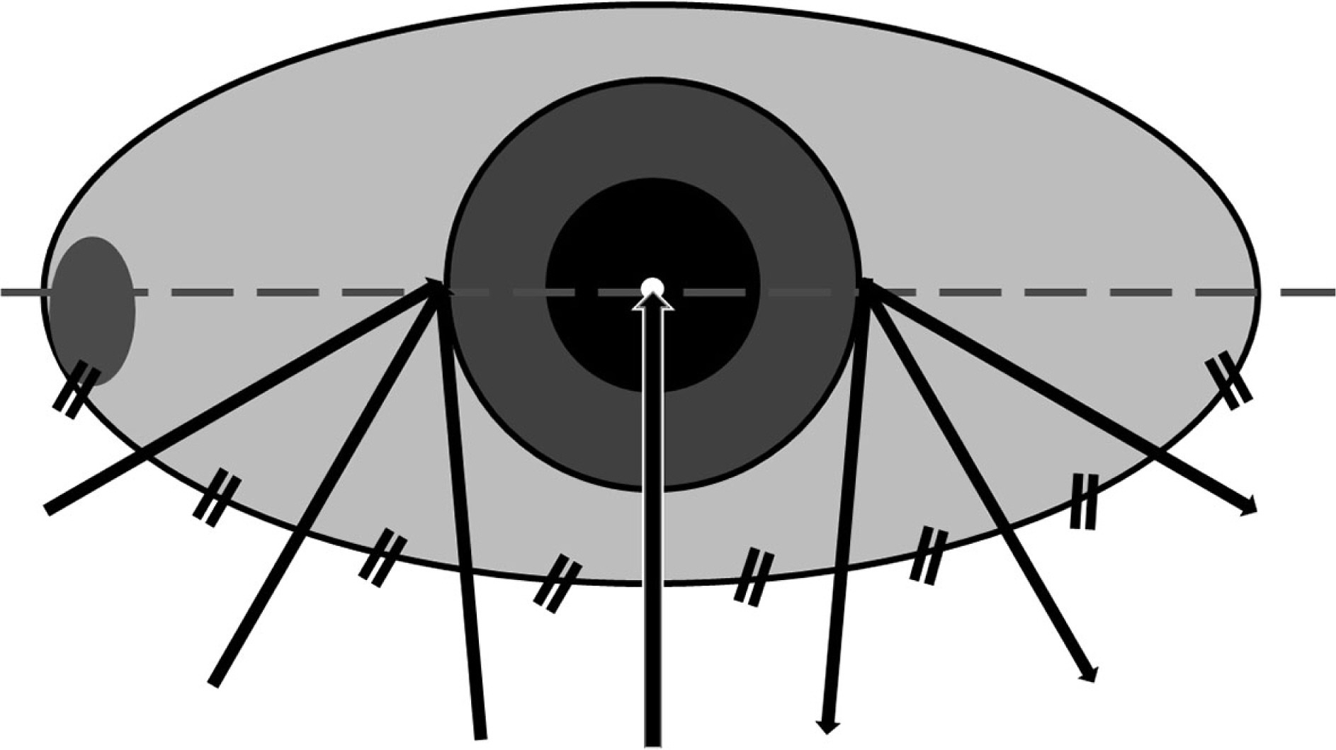
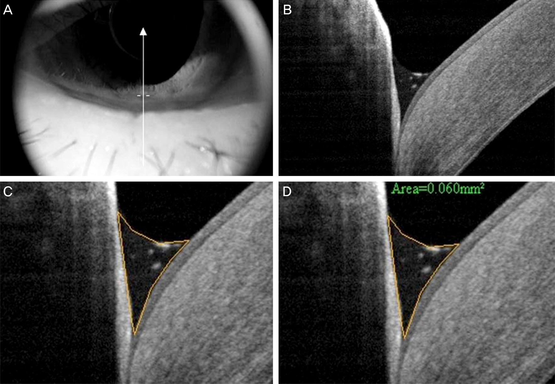
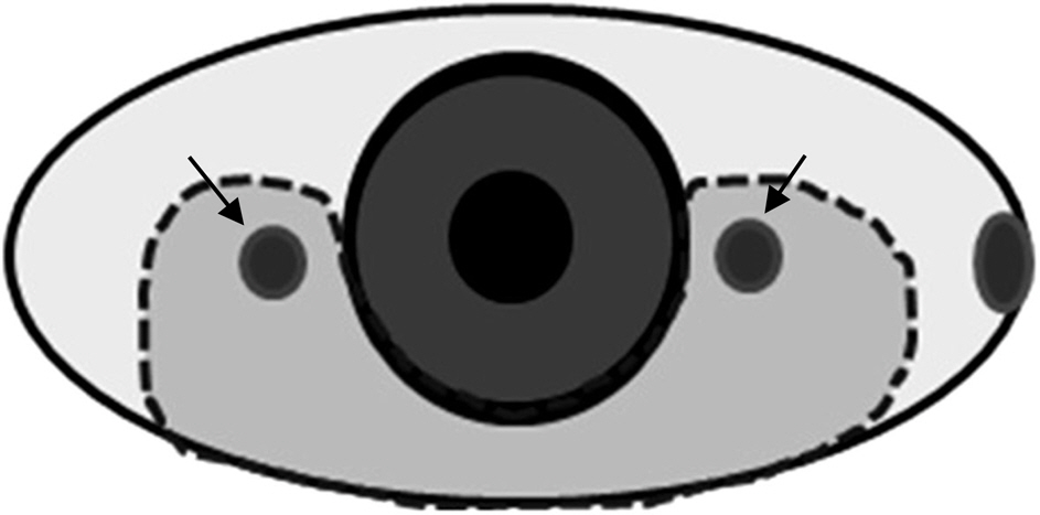
Thirty-one patients (33 eyes) with symptomatic conjunctivochalasis were recruited for this study between June 2013 and April 2014. All patients underwent crescent-shaped conjunctiva resection and amniotic membrane transplantation. Anterior segment OCT (AS-OCT) imaging was performed and tear break-up time was evaluated prior to and 3 months after the conjunctivochalasis surgery. Cross-sectional areas of conjunctiva and tear meniscus of conjunctivochalasis at 7 locations (1 center, 3 nasal and 3 temporal areas) were measured in all patients.
RESULTS
The mean age of patients was 66.3 +/- 10.8 years. Cross-sectional areas of conjunctivochalasis at all locations significantly decreased from 0.487 +/- 0.42 mm2 to 0.007 +/- 0.011 mm2 (p < 0.001), whereas no significant changes in cross-sectional areas of tear meniscus at all 7 locations were observed after the surgery. Mean tear break-up time significantly increased from 2.26 +/- 0.69 sec to 3.81 +/- 1.22 sec following the surgery.
CONCLUSIONS
Using AS-OCT, in this study we showed that areas of conjunctiva decreased and areas of tear meniscus were unchanged after conjunctivochalasis surgery.
Keyword
Figure
Cited by 1 articles
-
Combination Surgery of Silicone Tube Intubation and Conjunctival Resection in Patients with Epiphora
Seon Tae Kim, Long Yu Jin, Hee Bae Ahn
Korean J Ophthalmol. 2018;32(6):438-444. doi: 10.3341/kjo.2018.0044.
Reference
-
References
1. Meller D, Tseng SC. Conjunctivochalasis: literature review and possible pathophysiology. Surv Ophthalmol. 1998; 43:225–32.2. Wang Y, Dogru M, Matsumoto Y, et al. The impact of nasal conjunctivochalasis on tear functions and ocular surface findings. Am J Ophthalmol. 2007; 144:930–7.
Article3. Watanabe A, Yokoi N, Kinoshita S, et al. Clinicopathologic study of conjunctivochalasis. Cornea. 2004; 23:294–8.
Article4. Jordan DR, Pelletier CR. Conjunctivochalasis. Can J Ophthalmol. 1996; 31:192–3.5. Krachmer JH, Mannis MJ, Holland EJ. Cornea. 3rd ed.St. Louis: Mosby;2011. v. 1:p. 635–9.6. Hughes WL. Conjunctivochalasis. Am J Ophthalmol. 1942; 25:48–51.
Article7. Tseng SC, Prabhasawat P, Lee SH. Amniotic membrane transplantation for conjunctival surface reconstruction. Am J Ophthalmol. 1997; 124:765–74.
Article8. Meller D, Maskin SL, Pires RT, Tseng SC. Amniotic membrane transplantation for symptomatic conjunctivochalasis refractory to medical treatments. Cornea. 2000; 19:796–803.
Article9. Kheirkhah A, Casas V, Blanco G, et al. Amniotic membrane transplantation with fibrin glue for conjunctivochalasis. Am J Ophthalmol. 2007; 144:311–3.
Article10. Lim HJ, Lee JK, Park DJ. Conjunctivochalasis surgery: amniotic membrane transplantation with fibrin glue. J Korean Ophthalmol Soc. 2008; 49:195–204.
Article11. Nam K, Jo YJ, Lee SB. The efficacy of fibrin glue in surgical treatment of conjunctivochalasis with epiphora. J Korean Ophthalmol Soc. 2010; 51:498–503.
Article12. Oh SJ, Byon DS. Treatment of conjunctivochalasis using bipolar cautery. J Korean Ophthalmol Soc. 1999; 40:707–11.13. Youm DJ, Kim JM, Choi CY. Simple surgical approach with high-frequency radio-wave electrosurgery for conjunctivochalasis. Ophthalmology. 2010; 117:2129–33.
Article14. Shin KH, Hwang JH, Kwon JW. New approach for conjunctivochalasis with argon laser photocoagulation. Can J Ophthalmol. 2012; 47:380–2.
Article15. Huang D, Swanson EA, Lin CP, et al. Optical coherence tomography. Science. 1991; 254:1178–81.
Article16. Blumenthal EZ, Williams JM, Weinreb RN, et al. Reproducibility of nerve fiber layer thickness measurements by use of optical coherence tomography. Ophthalmology. 2000; 107:2278–82.17. Chen TC, Cense B, Pierce MC, et al. Spectral domain optical coherence tomography: ultra-high speed, ultra-high resolution ophthalmic imaging. Arch Ophthalmol. 2005; 123:1715–20.18. González-García AO, Vizzeri G, Bowd C, et al. Reproducibility of RTVue retinal nerve fiber layer thickness and optic disc measurements and agreement with Stratus optical coherence tomography measurements. Am J Ophthalmol. 2009; 147:1067–74. 1074.e1.
Article19. Menke MN, Dabov S, Knecht P, Sturm V. Reproducibility of retinal thickness measurements in patients with age-related macular degeneration using 3D Fourier-domain optical coherence tomography (OCT) (Topcon 3D-OCT 1000). Acta Ophthalmol. 2011; 89:346–51.20. Jittpoonkuson T, Garcia PM, Rosen RB. Correlation between fluorescein angiography and spectral-domain optical coherence tomography in the diagnosis of cystoid macular edema. Br J Ophthalmol. 2010; 94:1197–200.
Article21. Sakata LM, Lavanya R, Friedman DS, et al. Comparison of gonio-scopy and anterior segment ocular coherence tomography in de-tecting angle closure in different quadrants of the anterior chamber angle. Ophthalmology. 2008; 115:769–74.
Article22. Doors M, Tahzib NG, Eggink FA, et al. Use of anterior segment optical coherence tomography to study corneal changes after collagen cross-linking. Am J Ophthalmol. 2009; 148:844–51.e2.
Article23. Wang J, Aquavella J, Palakuru J, Chung S. Repeated measurements of dynamic tear distribution on the ocular surface after instillation of artificial tears. Invest Ophthalmol Vis Sci. 2006; 47:3325–9.
Article24. Chen Q, Wang J, Shen M, et al. Lower volumes of tear menisci in contact lens wearers with dry eye symptoms. Invest Ophthalmol Vis Sci. 2009; 50:3159–63.
Article25. Singh M, Aung T, Aquino MC, Chew PT. Utility of bleb imaging with anterior segment optical coherence tomography in clinical de-cision-making after trabeculectomy. J Glaucoma. 2009; 18:492–5.
Article26. Ciancaglini M, Carpineto P, Agnifili L, et al. Filtering bleb functionality: a clinical, anterior segment optical coherence tomography and in vivo confocal microscopy study. J Glaucoma. 2008; 17:308–17.
Article27. Gumus K, Crockett CH, Pflugfelder SC. Anterior segment optical coherence tomography: a diagnostic instrument for conjunctivochalasis. Am J Ophthalmol. 2010; 150:798–806.
Article28. Gumus K, Pflugfelder SC. Increasing prevalence and severity of conjunctivochalasis with aging detected by anterior segment optical coherence tomography. Am J Ophthalmol. 2013; 155:238–42.e2.
Article29. Zhou S, Li Y, Lu AT, et al. Reproducibility of tear meniscus measurement by Fourier-domain optical coherence tomography: a pilot study. Ophthalmic Surg Lasers Imaging. 2009; 40:442–7.
Article
- Full Text Links
- Actions
-
Cited
- CITED
-
- Close
- Share
- Similar articles
-
- Combination Surgery of Silicone Tube Intubation and Conjunctival Resection in Patients with Epiphora
- Tear Meniscus Evaluation Using Optical Coherence Tomography in Dry Eye Patients
- Changes in Tear Volume after 3% Diquafosol Treatment in Patients with Dry Eye Syndrome: An Anterior Segment Spectral-domain Optical Coherence Tomography Study
- Analysis of Tear Meniscus Change after Strabismus Surgery Using Optical Coherence Tomography
- Changes in Tear Meniscus Height Following Lower Blepharoplasty as Measured by Optical Coherence Tomography


