The Measurements of Macular Thickness and Volume with SD-OCT in Normal Eyes
- Affiliations
-
- 1Department of Ophthalmology, Ewha Womans University School of Medicine, Seoul, Korea. leejhoph@mm.ewha.ac.kr
- KMID: 2215000
- DOI: http://doi.org/10.3341/jkos.2011.52.10.1182
Abstract
- PURPOSE
We investigated reproducibility and repeatability of average macular thickness and volume measurements in normal eyes with Cirrus HD OCT (optical coherence tomography).
METHODS
Fifty normal eyes from twenty-five subjects without medical and ocular histories were included. Macular cube 512 x 128 combination scanning using the Cirrus HD OCT was performed for a total of three times on the same visit by an experienced technician. Then other two technicians performed one more macular scanning respectively. Within-results, the intraclass correlation coefficient (ICC) was calculated for each parameter studied to evaluate repeatability and reproducibility. The correlation between macular measurements and demographic variables (age, gender, and spherical equivalent) were also investigated.
RESULTS
The ICCs for intraoperator reproducibility were 0.99 on the average macular thickness and 0.96 on the macular volume. And the ICCs for interoperator repeatability were found to be 0.98 and 0.96, respectively. The ICCs for measurements of nine regional retinal thickness also were higher than 0.90. The retinal thickness was correlated with age, gender and refractive error. However, neither age nor refractive error affected to reproducibility and repeatability.
CONCLUSIONS
The retinal thickness and macular volume measurements using Cirrus HD OCT in healthy volunteers showed excellent reproducibility and repeatability. Therefore, Cirrus HD OCT has been recognized as an useful tool for diagnosis and mornitoring of variable maculopathies.
Keyword
Figure
-
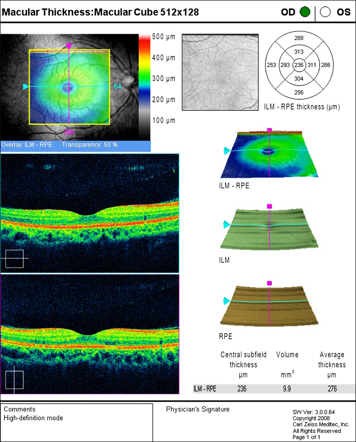
Figure 1. Standard output of Cirrus OCT, 512 × 128 macular cube (version 3.0.0.64, Carl Zeiss Meditec).
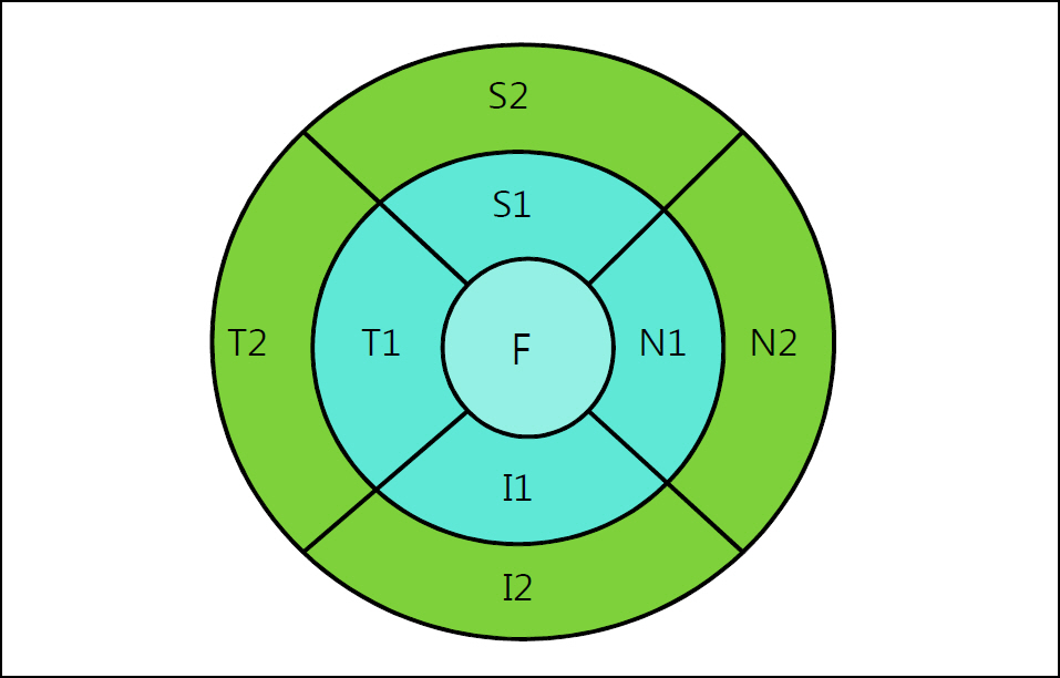
Figure 2. The macular thickness map of the right eye. F = fovea; S1 = superior inner; T1 = temporal inner; I1 = inferior inner; N1 = nasal inner; S2 = superior outer; T2 = temporal outer; I2 = inferior outer; N2 = nasal outer.
Cited by 3 articles
-
Foveal Shape According to Age and Gender Using Spectral Domain Optical Coherence Tomography
Min Byung Chae, Jae Suk Kim
J Korean Ophthalmol Soc. 2014;55(10):1504-1510. doi: 10.3341/jkos.2014.55.10.1504.A Study of Foveal Shape in Emmetropia and Myopia Using Spectral Domain Optical Coherence Tomography
Min Seok Kim, Jae Suk Kim, Jin Choi, Jung Hoon Kim, Won Hyuk Oh
J Korean Ophthalmol Soc. 2014;55(6):833-839. doi: 10.3341/jkos.2014.55.6.833.The Repeatability of Retinal Layer Thickness Measurements with Spectral-Domain Optical Coherence Tomography in Normal Eyes
Min Seok Kang, Seung-Young Yu, Hyung Woo Kwak
J Korean Ophthalmol Soc. 2016;57(5):786-793. doi: 10.3341/jkos.2016.57.5.786.
Reference
-
References
1. Huang J, Liu X, Wu Z, et al. Macular thickness measurements in normal eyes with time-domain and fourier-domain optical coherence tomography. Retina. 2009; 29:980–7.
Article2. Sayanagi K, Sharma S, Kaiser PK. Comparison of retinal thickness measurements between three-dimensional and radial scans on spectral-domain optical coherence tomography. Am J Ophthalmol. 2009; 148:431–8.
Article3. Hsu SY, Tung IC, Sheu MM, Tsai RK. Reproducibility of peripapillary retinal nerve fiber layer and macular retinal thickness measurements using optical coherence tomography. Kaohsiung J Med Sci. 2006; 22:447–51.
Article4. Ko BW, Shin YW, Lee JM, et al. Comparison of macular thickness measurements between Fourier-domain and Time-domain Optical coherence tomography in normal eyes and eyes with macular diseases. J Korean Ophthalmol Soc. 2009; 50:1661–8.
Article5. Ooto S, Hangai M, Sakamoto A, et al. Three-dimensional profile of macular retinal thickness in normal Japanese eyes. Invest Ophthalmol Vis Sci. 2010; 51:465–73.
Article6. Moon SW, Kim ES, Kim YG, et al. The comparison of macular thickness measurements and repeatabilities between time domain and spectral domain OCT. J Korean Ophthalmol Soc. 2009; 50:1050–9.
Article7. Wong AC, Chan CW, Hui SP. Relationship of gender, body mass index, and axial length with central retinal thickness using optical coherence tomography. Eye. 2005; 19:292–7.
Article8. Oh SB, Cho WB, Moon JW, Kim HC. Repeatability and agreement of macular thickness measurement using time domain OCT and spectral domain OCT in normal subjects. J Korean Ophthalmol Soc. 2009; 50:710–6.
Article9. Menke MN, Dabov S, Knecht P, Sturm V. Reproducibility of retinal thickness measurements in healthy subjects using spectralis optical coherence tomography. Am J Ophthalmol. 2009; 147:467–72.
Article10. Leung CK, Cheung CY, Weinreb RN, et al. Comparison of macular thickness measurements between time domain- and spectral domain-optical coherence tomography (OCT). Invest Ophthalmol Vis Sci. 2008; 49:4893–7.11. Han IC, Jaffe GJ. Comparison of spectral- and aberrations optical coherence tomography for retinal thickness measurements in healthy and diseased eyes. Am J Ophthalmol. 2009; 147:847–58.12. Kiernan DF, Mieler WF, Hariprasad SM. Spectral-domain optical coherence tomography: a comparison of modern high-resolution retinal imaging systems. Am J Ophthalmol. 2010; 149:18–31.
Article13. Kiernan DF, Hariprasad SM, Chin EK, et al. Prospective comparison of cirrus and stratus optical coherence tomography for quantifying retinal thickness. Am J Ophthalmol. 2009; 147:267–75.
Article14. Early Treatment Diabetic Retinopathy Study Research Group. Photocoagulation for diabetic macular edema. Early Treatment Diabetic Retinopathy Study report number 1. Arch Ophthalmol. 1985; 103:1796–806.15. Maldonado MJ, Nieto JC, Diez-Cuenca M, Pinero DP. Repeatability and reproducibility of posterior corneal curvature measurements by combined scanning-slit and placido-disc topography after LASIK. Ophthalmology. 2006; 113:1918–26.
Article16. Muscat S, Parks S, Kemp E, Keating D. Repeatability and reproducibility of macular thickness measurements with the Humphrey OCT system. Invest Ophthalmol Vis Sci. 2002; 43:490–5.17. Syc SB, Warner CV, Hiremath GS, et al. Reproducibility of aberrations optical coherence tomography in multiple sclerosis. Mult Scler. 2010; 16:829–39.18. Müller R, Büttner P. A critical discussion of intraclass correlation coefficients. Stat Med. 1994; 13:2465–76.
Article19. Polito A, Shah SM, Haller JA, et al. Comparison between retinal thickness analyzer and optical coherence tomography (OCT) for assessment of foveal thickness in eyes with macular disease. Am J Ophthalmol. 2002; 134:240–51.20. Legarreta JE, Gregori G, Punjabi OS, et al. Macular thickness measurements in normal eyes using spectral domain optical coherence tomography. Ophthalmic Surg Lasers Imaging. 2008; 39:s43–9.
Article21. Kanai K, Abe T, Murayama K, Yoneya S. Retinal thickness and changes with age. Nippon Ganka Gakkai Zasshi. 2002; 106:162–5.
Article22. Guedes V, Schuman JS, Hertzmark E, et al. Optical coherence tomography measurement of macular and nerve fiber layer thickness in normal and glaucomatous human eyes. Ophthalmology. 2003; 110:177–89.
Article23. Panda-Jonas S, Jonas JB, Jakobczyk-Zmija M. Retinal photoreceptor density decreases with age. Ophthalmology. 1995; 102:1853–9.
Article24. Kashani AH, Zimmer-Galler IE, Shah SM, et al. Retinal thickness analysis by race, gender, and age using stratus OCT. Am J Ophthalmol. 2010; 149:496–502.
Article25. Shin JH, Lee HJ, Jin KH. The relationship between axial length, refractive power and foveal thickness measured by OCT in Koreans. J Korean Ophthalmol Soc. 2005; 46:701–6.26. Massin P, Erginay A, Haouchine B, et al. Retinal thickness in healthy and diabetic subjects measured using optical coherence tomography mapping software. Eur J Ophthalmol. 2002; 12:102–8.
Article27. Salyer DL, Lund TD, Fleming DE, et al. Sexual dimorphism and aromatase in the rat retina. Brain Res Dev Brain Res. 2001; 126:131–6.
Article28. Keane PA, Mand PS, Liakopoulos S, et al. Accuracy of retinal thickness measurements obtained with Cirrus optical coherence tomography. Br J Ophthalmol. 2009; 93:1461–7.
Article
- Full Text Links
-
- Actions
-
Cited
- CITED
-
- Close
- Share
-
- Similar articles
-
- Repeatability and Agreement of Macular Thickness Measurement Using Time and Spectral Domain OCT in Diabetic Macular Edema
- Repeatability and Agreement of Macular Thickness Measurement Using Time Domain OCT and Spectral Domain OCT in Normal Subjects
- The Comparison of Macular Thickness Measurements and Repeatabilities Between Time Domain and Spectral Domain OCT
- Comparison of Macular Thickness Measurements Between Fourier-Domain and Time-Domain Optical Coherence Tomography in Normal Eyes and Eyes With Macular Diseases
- Repeatability of Spectral Domain OCT (3D-OCT 1000) in Normal Subjects and Various Macular Diseases
