Bilateral Coats' Disease: A Case Report
- Affiliations
-
- 1Department of Ophthalmology, Seoul National University College of Medicine, Seoul, Korea. ysyu@snu.ac.kr
- 2Seoul Artificial Eye Center, Seoul National University Hospital Clinical Research Institute, Seoul, Korea.
- KMID: 2214147
- DOI: http://doi.org/10.3341/jkos.2011.52.1.112
Abstract
- PURPOSE
To report a case of bilateral Coats' disease.
CASE SUMMARY
A 19-month-old boy presented with esodeviation of his eyes, which started 5 months prior. A fundus exam showed total bullous exudative retinal detachment with retinal vascular telangiectasia in the right eye and localized exudative retinal detachment with vascular telangiectasia at the inferior periphery in the right eye. Fluorescein angiogram of the left eye showed retinal telangiectatic vessels, avascular area and fluorescein leakeage from telangiectatic vessels. The patient received external drainage of subretinal fluid and intravitreal air injection of the right eye and Argon LASER photocoagulation and cryotheraphy of the left eye. A cytologic exam of the subretinal fluid drained from the right eye showed no malignant cells. Forty-four months after the operation, his best corrected visual acuity was no light perception in the right eye and 0.4 in the left eye. Both fundi were flat and stable. No complications, such as glaucoma, recurred retinal detachment, or pain, occurred.
CONCLUSIONS
Coats' disease rarely occurs bilaterally and can be involved asymmetrically. The disease presents more severely when bilateral and can progress after long-term observation. Proper treatment and long-term follow-up of both eyes are necessary to prevent visual loss and preserve eyes.
MeSH Terms
Figure
-
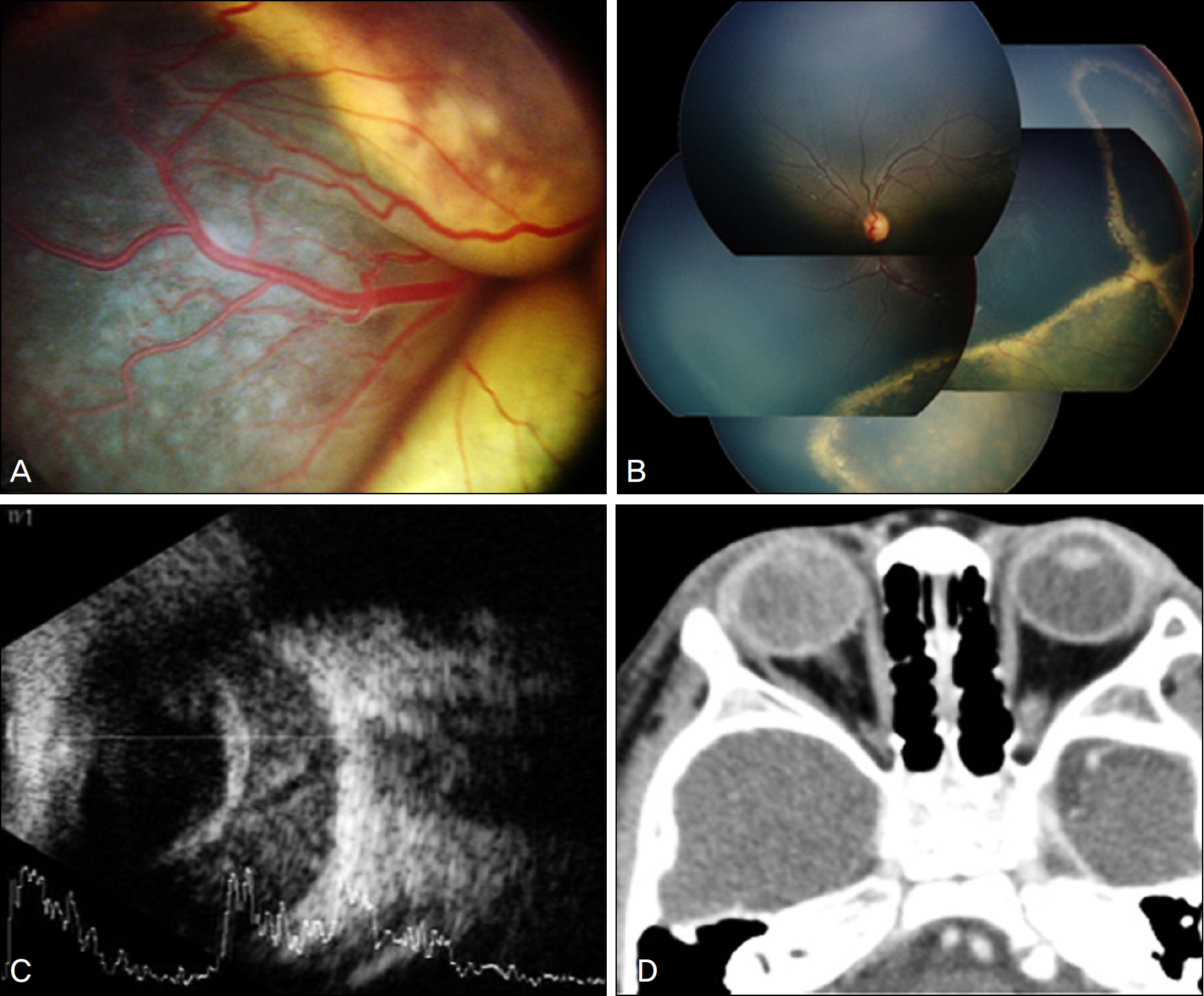
Figure 1. Initial examination findings. (A) Total bullous exudative retinal detatchment with retinal vascular telangiectasia in the right eye. (B) Fundus photograph showing characteristic features of Coats' disease with localized exudative retinal detachment with vascular telangiectasia at the inferior periphery in the left eye. (C) Ocular ultrasonography showing no mass-like lesion. (D) Orbit CT showing no mass-like lesion.
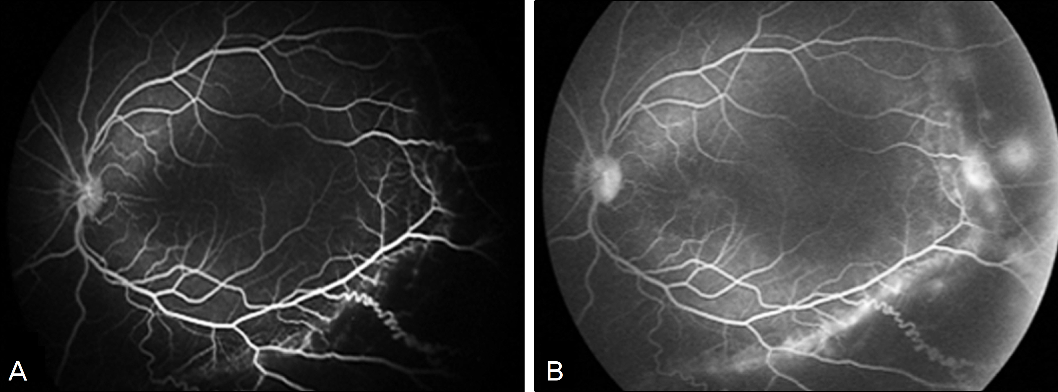
Figure 2. Fluorescein angiogram of the left eye before the laser photocoagulation. (A) Retinal telangiectatic vessels and avascular area. (B) Fluorescein leakeage from telangiectatic vessels.
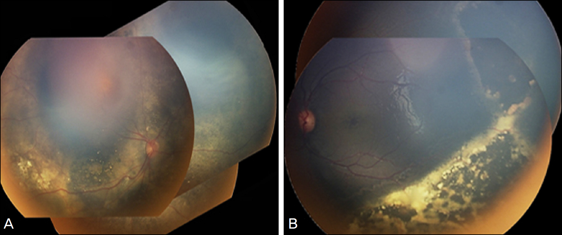
Figure 3. Fundus photograph taken 2 months after operation. (A) Right eye, flat. (B) Left eye, subretinal fluid decreased a great deal.
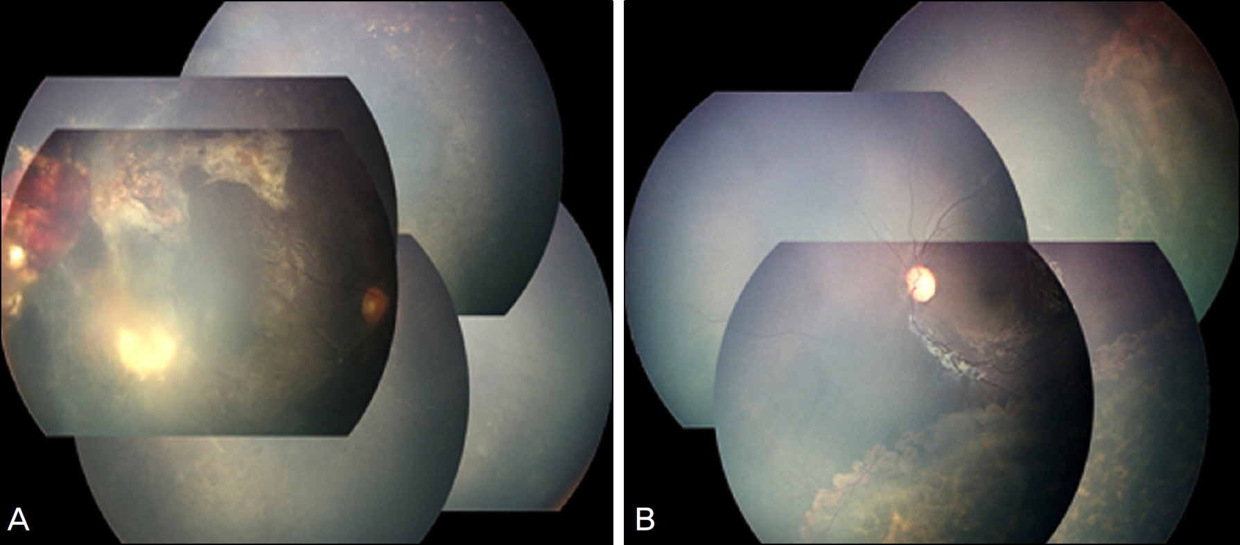
Figure 4. Fundus photograph taken 33 months after operation. (A) Right eye. (B) Left eye. Both fundi are flat and stable.
Cited by 2 articles
-
Letter to the Editor: Bilateral Coats' Disease: A Case Report
Yu Cheol Kim
J Korean Ophthalmol Soc. 2011;52(5):644-645. doi: 10.3341/jkos.2011.52.5.644.Clinical Characteristics of Retinoblastoma Patients whose Diagnosis was Difficult due to Atypical Ocular Manifestation
Haeng Jin Lee, Dong Hyun Jo, Jeong Hun Kim, Young Suk Yu
J Korean Ophthalmol Soc. 2016;57(5):829-836. doi: 10.3341/jkos.2016.57.5.829.
Reference
-
References
1. Coats G. Forms of retinal diseases with massive exudation. Roy Lond Ophthalmol Hosp Rep. 1908; 17:440–525.2. Leber T. Ueber eine durch Vorkommen multipler Miliaraneurysmen charakterisierte Form von Retinaldegeneration. Albrecht von Graefes Arch Ophthalmol. 1912; 81:1–14.3. Reese AB. Telangiectasis of the retina and Coats' disease. Am J Ophthalmol. 1956; 42:1–8.
Article4. Green WR. Bilateral Coats' disease. Massive gliosis of the retina. Arch Ophthalmol. 1967; 77:378–83.5. McGettrick PM, Loeffler KU. Bilateral Coats' disease in an infant (a clinical, angiographic, light and electron microscopic study). Eye. 1987; 1:136–45.
Article6. Alexandridou A, Stavrou P. Bilateral Coats' disease: long-term follow up. Acta Ophthalmol Scand. 2002; 80:98–100.
Article7. Shields JA, Shields CL, Honavar S, Demirci H. Coats disease. Clinical variations and complications of Coats disease in 150 cases. The 2000 Sanford Gifford Memorial Lecture. Am J Ophthalmol. 2001; 131:561–71.8. Woods AC, Duke JR. Coats's disease. I. Review of the literature diagnostic criteria, clinical findings, and plasma lipid studies. Br J Ophthalmol. 1963; 47:385–412.
Article9. Gomez Morales A. Coats' disease. Natural history and results of treatment. Am J Ophthalmol. 1965; 60:855–65.10. Egerer I, Tasman W, Tomer TT. Coats disease. Arch Ophthalmol. 1974; 92:109–12.
Article11. Tarkkanen A, Laatikainen L. Coat's disease: clinical, angiographic, histopathological findings and clinical management. Br J aberrations. 1983; 67:766–76.
Article12. Chang MM, McLean IW, Merritt JC. Coats' disease: a study of 62 histologically confirmed cases. J Pediatr Ophthalmol Strabismus. 1984; 21:163–8.
Article13. Silodor SW, Augsburger JJ, Shields JA, Tasman W. Natural history and management of advanced Coats' disease. Ophthalmic Surg. 1988; 19:89–93.
Article14. Shields JA, Shields CL, Honavar SG, et al. Classification and management of Coats disease. The 2000 Proctor Lecture. Am J Ophthalmol. 2001; 131:572–83.
Article15. Shields JA, Shields CL. Review: Coats disease: the 2001 LuEsther T. Mertz lecture. Retina. 2002; 22:80–91.16. Shienbaum G, Tasman WS. Coats disease: a lifetime disease. Retina. 2006; 26:422–4.17. Rishi P, Rishi E, Uparkar M, et al. Coats' disease: an Indian perspective. Indian J Ophthalmol. 2010; 58:119–24.
Article18. Lai CH, Kuo HK, Wu PC, et al. Manifestation of Coats' disease by age in Taiwan. Clin Experiment Ophthalmol. 2007; 35:361–5.
Article19. Kim WS, Jung BJ, Kim JW, et al. A Case of Coats' disease. J Korean Ophthalmol Soc. 1969; 10:37–9.20. Choi SY, Yu YS. Treatment and clinical results of coats' disease. J Korean Ophthalmol Soc. 1999; 40:2190–7.21. Han ES, Choung HK, Heo JW, et al. The Clinical analysis and treatment results of Coats' disease in children. J Korean Ophthalmol Soc. 2006; 47:423–30.
- Full Text Links
-
- Actions
-
Cited
- CITED
-
- Close
- Share
-
- Similar articles
-
- A Case of Retinoblastoma and Coats' Disease in the Same eye: A Clinicopathologic Report
- Fluorescein Angiographic Abnormalities in the Contralateral Eye with Normal Fundus in Children with Unilateral Coats' Disease
- Letter to the Editor: Bilateral Coats' Disease: A Case Report
- Intravitreal Ranibizumab Injection in Adult-onset Coats' Disease: A Case Report
- A Case of Coats' Disease With Spontaneous Peeling of Premacular Membrane After Photocoagulation
