Comparison of Effective Phacoemulsification Time between Femtosecond Laser-Assisted Cataract Surgery and Conventional Cataract Surgery
- Affiliations
-
- 1Sungmo Eye Hospital, Busan, Korea. medicalhan@hanmail.net
- KMID: 2213664
- DOI: http://doi.org/10.3341/jkos.2016.57.2.236
Abstract
- PURPOSE
To compare the effect of femtosecond laser-assisted cataract surgery with conventional cataract surgery on effective phacoemulsification time (EPT).
METHODS
This study included 66 patients 100 eyes who underwent femtosecond laser-assisted cataract surgery and 68 patients 100 eyes who underwent conventional cataract surgery. Both groups underwent phacoemulsification using pulsed ultrasound energy and EPT was evaluated. The groups were further analyzed according to preoperative Lens opacities classification system (LOCS) III grading. Patients who had femtosecond laser-assisted cataract surgery underwent lens fragmentation with quadrant, hybrid, or grid pattern and the EPT was respectively evaluated.
RESULTS
The mean EPT was 5.85 +/- 4.31 seconds in the femtosecond laser-assisted cataract surgery group and 10.34 +/- 6.61 seconds in the conventional group. Overall, EPT was statistically significantly lower in the femtosecond laser-assisted cataract surgery group compared to the conventional group. When the groups were analyzed according to LOCS III grading, this result was consistent for all cataract grades and the reduction in EPT was increased with the higher LOCS III grade. When the groups were analyzed according to lens fragmentation patterns, the mean EPT was lower with 350 microm grid pattern than the quadrant or hybrid pattern.
CONCLUSIONS
The femtosecond laser-assisted system in cataract surgery can be an efficient cataract surgery using lower EPT compared to the conventional procedure. Additionally, significant differences were observed in the mean EPT of cataract surgery using the femtosecond laser-assisted system among the 3 lens fragmentation pattern groups.
Figure
-
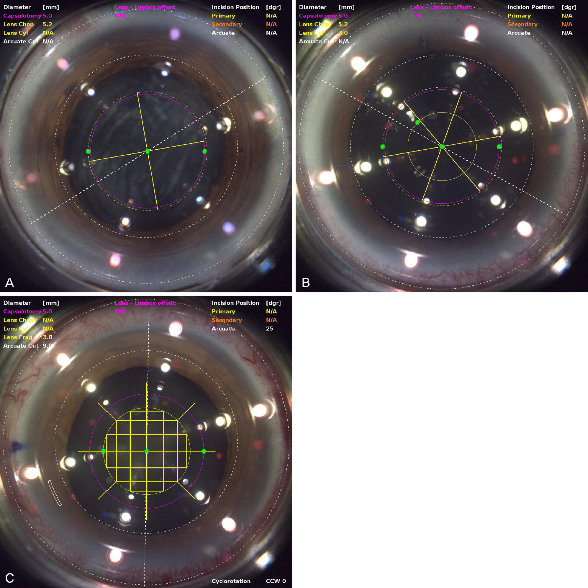
Figure 1. Fragmentation pattern during cataract surgery. (A) Quadrant pattern, (B) hybrid pattern, and (C) grid pattern. LenSx real-time imaging showing planned capsulotomy (pink circle) and lens fragmentation (yellow line). N/A = not avail-able; cyl = cylinder; dgr = degree; CCW = counterclockwise.
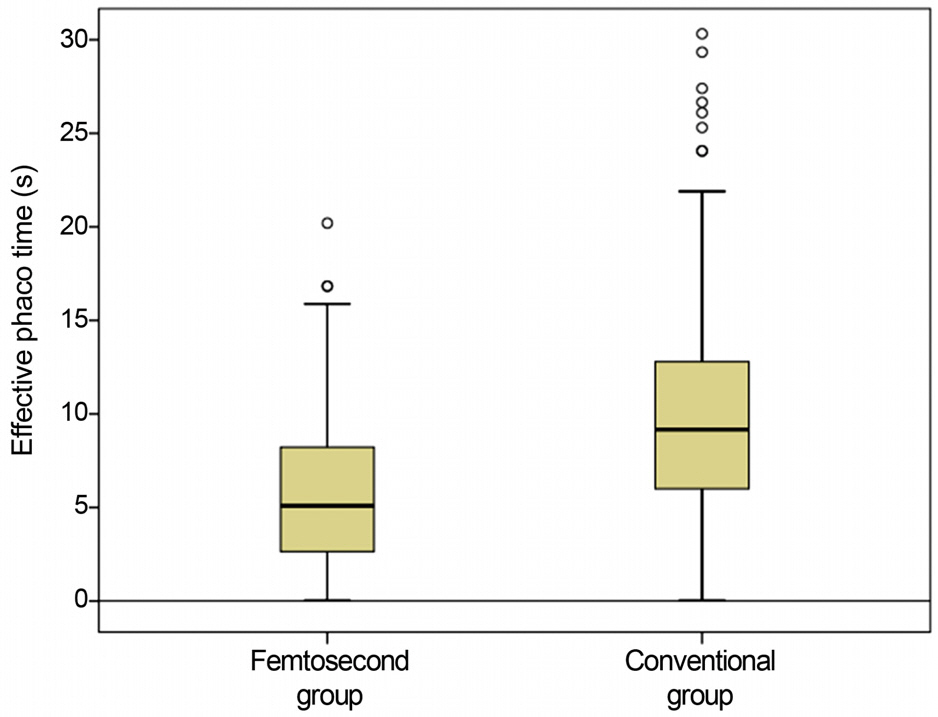
Figure 2. Effective phacoemulsification time comparison between femtosecond laser-assisted cataract surgery (femtosecond) group and conventional cataract surgery group. Student's t-test was used to examine statistical difference.

Figure 3. Effective phacoemulsification time in the femtosecond group and conventional group in regard to the nuclear opacity (LOCS III). Student's t-test was used to examine statistical difference. NO = nuclear opacity by LOCS III classification.
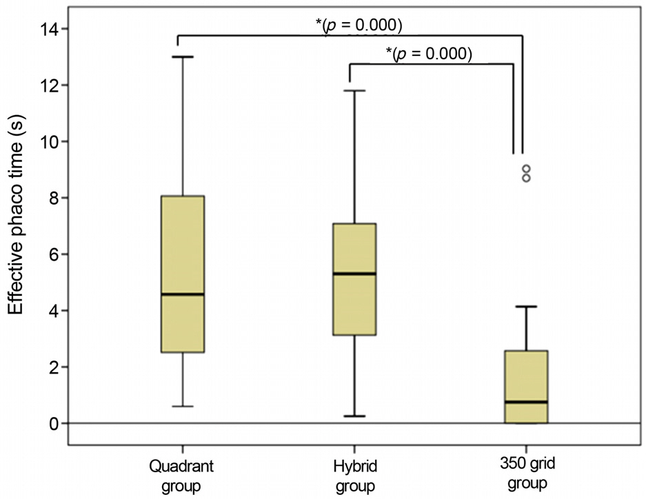
Figure 4. Effective Phacoemulsification Time comparison of different lens fragmentation in the femtosecond group. Kruskal-Wallis test was used to examine statistical difference. * Significant difference between groups.
Cited by 1 articles
-
Long-term Results of Arcuate Keratotomy in Femtosecond Laser-assisted Cataract Surgery
Chan Woo Bang, Jae Won Choi, Sang Youp Han
J Korean Ophthalmol Soc. 2019;60(10):946-952. doi: 10.3341/jkos.2019.60.10.946.
Reference
-
1). Sugar A. Ultrafast (femtosecond) laser refractive surgery. Curr Opin Ophthalmol. 2002; 13:246–9.
Article2). Juhasz T, Loesel FH, Kurtz RM, et al. Corneal refractive surgery with femtosecond lasers. IEEE Journal of Selected Topics in Quantum Electronics. 1999; 5:902–10.
Article3). Kohnen T, Klaproth OK, Derhartunian V, Kook D. Results of 308 consecutive femtosecond laser cuts for LASIK. Ophthalmologe. 2010; 107:439–45.4). Chang JS, Chen IN, Chan WM, et al. Initial evaluation of a femtosecond laser system in cataract surgery. J Cataract Refract Surg. 2014; 40:29–36.
Article5). Park JH, Lee KH, Lee DJ. Comparison of continuous curvilinear capsulorhexis parameters between femtosecond laser and conventional cataract surgery. J Korean Ophthalmol Soc. 2014; 55:1800–7.
Article6). Masket S, Sarayba M, Ignacio T, Fram N. Femtosecond laser-assisted cataract incisions: architectural stability and reproducibility. J Cataract Refract Surg. 2010; 36:1048–49.
Article7). Mayer WJ, Klaproth OK, Hengerer FH, Kohnen T. Impact of crystalline lens opacification on effective phacoemulsification time in femtosecond laser-assisted cataract surgery. Am J Ophthalmol. 2014; 157:426–32. e1.
Article8). Nagy ZZ, Ecsedy M, Kovács I, et al. Macular morphology assessed by optical coherence tomography image segmentation after femtosecond laser-assisted and standard cataract surgery. J Cataract Refract Surg. 2012; 38:941–6.
Article9). Bencić G, Zorić-Geber M, Sarić D, et al. Clinical importance of the lens opacities classification system III (LOCS III) in phacoemulsification. Coll Antropol. 2005; 29:Suppl 1. 91–4.10). Richard J, Hoffart L, Chavane F, et al. Corneal endothelial cell loss after cataract extraction by using ultrasound phacoemulsification versus a fluid-based system. Cornea. 2008; 27:17–21.
Article11). Hayashi K, Hayashi H, Nakao F, Hayashi F. Risk factors for corneal endothelial injury during phacoemulsification. J Cataract Refract Surg. 1996; 22:1079–84.
Article12). Werblin TP. Long-term endothelial cell loss following phacoemulsification: model for evaluating endothelial damage after intraocular surgery. Refract Corneal Surg. 1993; 9:29–35.
Article13). Nagy Z, Takacs A, Filkorn T, Sarayba M. Initial clinical evaluation of an intraocular femtosecond laser in cataract surgery. J Refract Surg. 2009; 25:1053–60.
Article14). Palanker DV, Blumenkranz MS, Andersen D, et al. Femtosecond laser-assisted cataract surgery with integrated optical coherence tomography. Sci Transl Med. 2010; 2:58ra85.
Article15). Conrad-Hengerer I, Hengerer FH, Schultz T, Dick HB. Effect of femtosecond laser fragmentation on effective phacoemulsification time in cataract surgery. J Refract Surg. 2012; 28:879–83.
Article16). Abell RG, Kerr NM, Vote BJ. Femtosecond laser-assisted cataract surgery compared with conventional cataract surgery. Clin Experiment Ophthalmol. 2013; 41:455–62.
Article17). Lee WS, Han SY, Lee KH. Comparison of laser refractive cataract surgery with a femtosecond laser versus conventional phacoemulsification. J Korean Ophthalmol Soc. 2013; 54:1227–35.
Article18). Conrad-Hengerer I, Hengerer FH, Schultz T, Dick HB. Effect of femtosecond laser fragmentation of the nucleus with different softening grid sizes on effective phaco time in cataract surgery. J Cataract Refract Surg. 2012; 38:1888–94.
Article19). Roberts TV, Sutton G, Lawless MA, et al. Capsular block syndrome associated with femtosecond laser-assisted cataract surgery. J Cataract Refract Surg. 2011; 37:2068–70.
Article20). Murano N, Ishizaki M, Sato S, et al. Corneal endothelial cell damage by free radicals associated with ultrasound oscillation. Arch Ophthalmol. 2008; 126:816–21.
Article21). Shin YJ, Nishi Y, Engler C, et al. The effect of phacoemulsification energy on the redox state of cultured human corneal endothelial cells. Arch Ophthalmol. 2009; 127:435–41.
Article22). Kohnen T. Compromised corneal endothelium and cataract: how should we decide? J Cataract Refract Surg. 2011; 37:1377–8.
Article23). Kim DS, Kim MJ, Kim JY, Tchah HW. Comparison of the phaco chop and the mini chop methods in cataract surgery. J Korean Ophthalmol Soc. 2005; 46:1780–5.24). Abell RG, Kerr NM, Vote BJ. Toward zero effective phacoemulsification time using femtosecond laser pretreatment. Ophthalmology. 2013; 120:942–8.
Article
- Full Text Links
-
- Actions
-
Cited
- CITED
-
- Close
- Share
-
- Similar articles
-
- Short-term Clinical Outcomes of Femtosecond Laser-assisted Cataract Surgery: Comparison with Conventional Phacoemulsification
- Comparison of Laser Refractive Cataract Surgery with a Femtosecond Laser Versus Conventional Phacoemulsification
- Comparison of Continuous Curvilinear Capsulorhexis Parameters between Femtosecond Laser and Conventional Cataract Surgery
- Long-term Results of Arcuate Keratotomy in Femtosecond Laser-assisted Cataract Surgery
- Comparative Study of Microcoaxial Cataract Surgery and Conventional Cataract Surgery
