J Korean Ophthalmol Soc.
2010 Apr;51(4):611-615. 10.3341/jkos.2010.51.4.611.
A Case of Keratoconjunctivitis Caused by Chlamydophila Psittaci
- Affiliations
-
- 1Department of Ophthalmology, Ewha Womans University School of Medicine, Seoul, Korea. jrmoph@ewha.ac.kr
- KMID: 2213407
- DOI: http://doi.org/10.3341/jkos.2010.51.4.611
Abstract
- PURPOSE
Only a few cases of keratoconjunctivitis caused by Chlamydophila psittaci have been reported worldwide, and no case reported in Korea. We report an atypical case of keratoconjunctivitis caused by Chlamydophila psittaci.
CASE SUMMARY
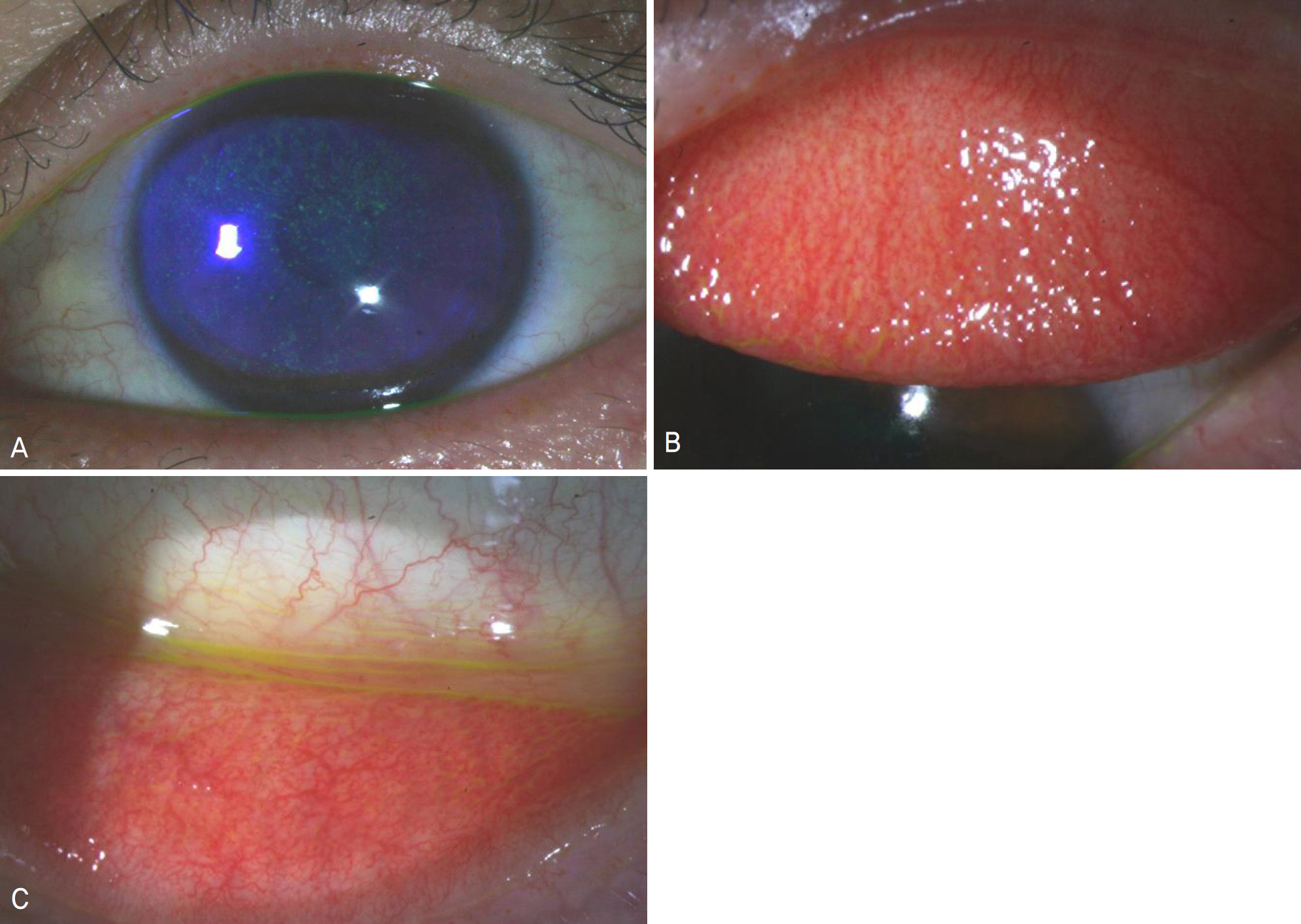
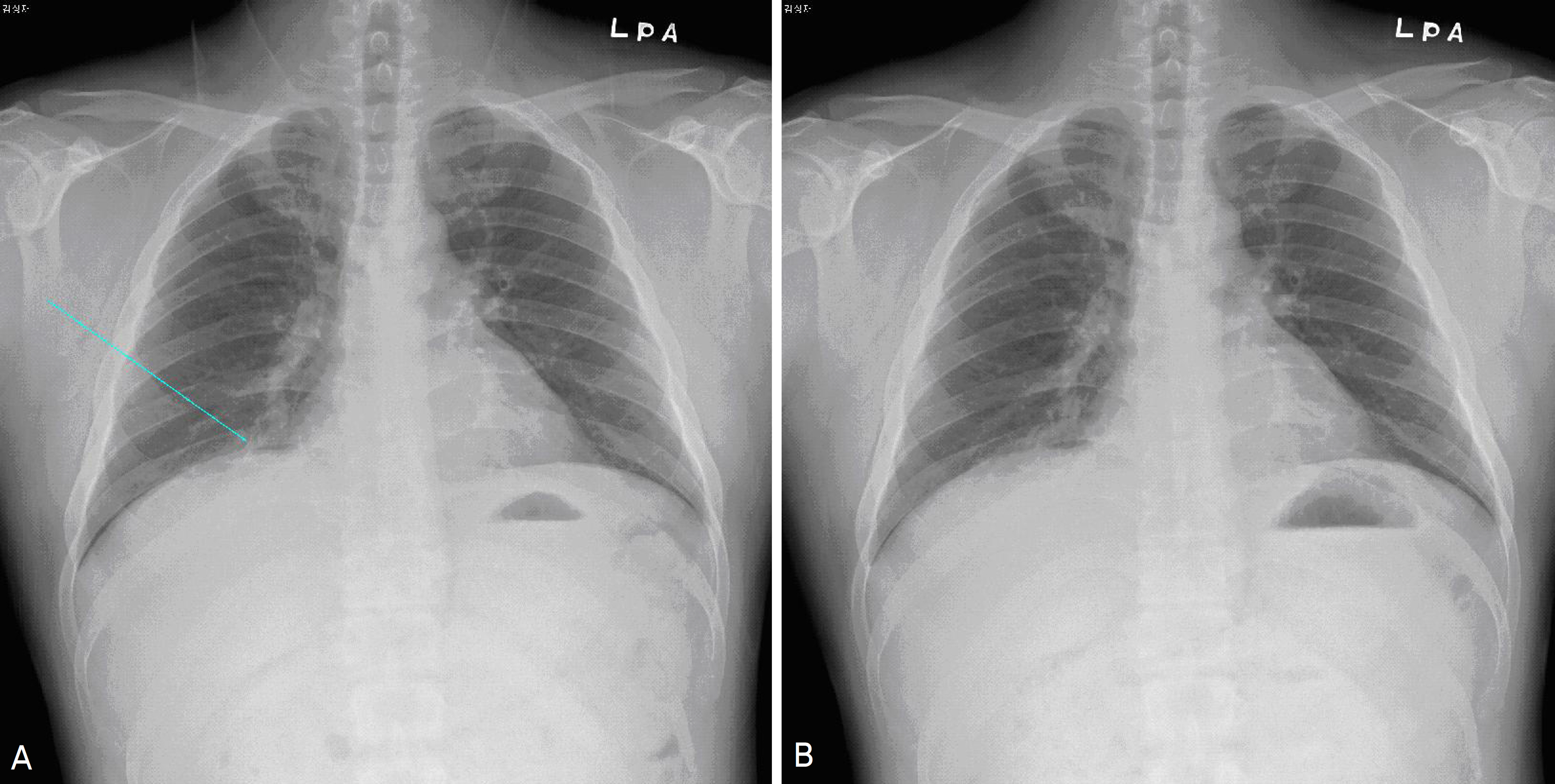
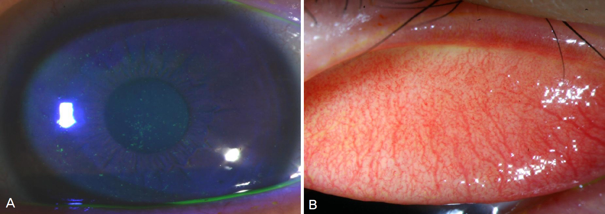
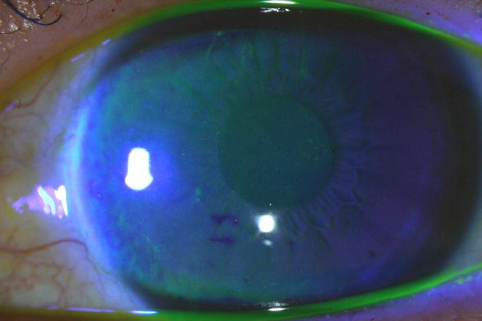
A 34-year-old male patient who had raised a parrot at home presented with three weeks of conjunctival injection and a week of ocular pain in his left eye. There were papillae on the left upper and lower tarsal conjunctiva and punctuate epithelial erosion of the entire cornea. He also complained of dizziness, fever, and dyspnea. Upon chest X-ray, consolidation on the right middle lobe was apparent. The Chlamydophila IgM antibody test was positive, and the pneumonia improved quickly. Nevertheless, signs of keratoconjunctivitis persisted despite 3-week treatment with oral doxycycline. As a result, the patient received an additional 10-day treatment with oral azithromycin. Four weeks after the first visit, symptoms were improving gradually, and, after six weeks, no signs of keratoconjunctivitis remained except minimal erosion.
CONCLUSIONS
When patients show keratoconjunctivitis after contact with a bird, prolonged ketatoconjunctivitis by Chlamydophila psittaci should be considered.
Keyword
MeSH Terms
Figure
Reference
-
References
1. Dean D, Shama A, Schachter J, Dawson CR. Molecular abdominal of an avian strain of Chlamydia psittaci causing severe keratoconjunctivitis in a bird fancier. Clin Infect Dis. 1995; 20:1179–85.2. Krachmer JH, Mannis MJ, Holland EI. Cornea. 2nd ed.Vol. 1. Philadelphia: Elsevier Inc;2005. p. 639–46.3. Ostler HB, Schachter J, Dawson CR. Acute follicular abdominal of epizootic origin. Arch Ophthalmol. 1969; 82:587–91.4. Schachter J, Ostler HB, Meyer KF. Human infection with the agent of feline pneumonitis. Lancet. 1969; 1:1063–5.
Article5. Lietma T, Brooks D, Moncada J, et al. Chronic follicular abdominal associated with Chlamydia psittaci or Chlamydia pneumoniae. Clin Infect Dis. 1998; 26:1335–40.6. Schacter J, Arnstein P, Dawson CR, et al. Human follicular abdominal caused by infection with a psittacosis agent. Proc Soc Exp Biol Med. 1968; 127:292–5.7. Niki Y, Kimura M, Miyashita N, Soejima R. In vitro and in vivo activities of azithromycin, a new azalide antibiotic, against chlamydia. Antimicrob Agents Chemother. 1994; 38:2296–9.
Article8. Donati M, Rodrìguez Fermepin M, Olmo A, et al. Comparative in-vitro activity of moxifloxacin, minocycline and azithromycin against chlamydia spp. J Antimicrob Chemother. 1999; 43:825–7.
Article
- Full Text Links
- Actions
-
Cited
- CITED
-
- Close
- Share
- Similar articles
-
- Clinico-pathological Features of Chlamydophila psittaci Infection in Parrots and Genetic Characterization of the Isolates
- Detection of Chlamydophila pneumoniae in Acute Myocardial Infarction
- Detection of Chlamydia pneumoniae by 'Touchdown' PCR
- A Case of Gonococcal Keratoconjunctivitis in Adult
- Multiplex Real-time PCR Assay for the Detection of all Chlamydia Species and Simultaneous Differentiation of C. psittaci and C. pneumoniae in Human Clinical Specimens


