Electrophysiological and Morphological Changes After Intravitreal Bevacizumab Injection with Macular Edema or Choroidal Neovascularization
- Affiliations
-
- 1Department of Ophthalmology, Soonchunhyang University College of Medicine, Cheonan, Korea.
- 2Department of Ophthalmology, Soonchunhyang University College of Medicine, Bacheon, Korea. yhohn@schbc.ac.kr
- KMID: 2212994
- DOI: http://doi.org/10.3341/jkos.2009.50.12.1824
Abstract
- PURPOSE
To evaluate the functional and anatomical statuses of macula with multifocal electroretinogram (mfERG) and optical coherence tomography (OCT) for the treatment of patients with macular edema after intravitreal bevacizumab injection.
METHODS
Patients were injected with intravitreal bevacizumab (1.875 mg/0.075 ml) for macular edema with choroidal neovascularization (CNV), diabetic retinopathy and retinal vein occlusion.
RESULTS
A total of 120 eyes (96 patients) were diagnosed with diabetic retinopathy, CNV with age-related macular degeneration and retinal vein occlusion. Visual acuity improved from logMAR 0.85+/-0.39 to 0.54+/-0.38 after intravitreal bevacizumab injection. The implicit time of P1 and N2 in mfERG decreased, and the amplitude of P1 showed a statistically significant increase. Central macular thickness decreased from 374.4+/-135.2 micrometer to 249.0+/-72.0 micrometer.
CONCLUSIONS
After intravitreal bevacizumab injection, functional and anatomical statuses of mfERG and OCT improved. This study demonstrates a method for utilizing mfERG to assess the effectiveness of treatments such as bevacizumab.
Keyword
MeSH Terms
Figure
-

Figure 1. Changes in plot of mfERG after intravitreal bevacizumab injection; implicit time was shortened and retinal response densities were increased on ring 1 and 2 areas of mfERG after IVB injection. Peak of retinal response densities of macular area were recovered on 3-D plot after IVB injection.* IVB=intravitreal bevacizumab injection.
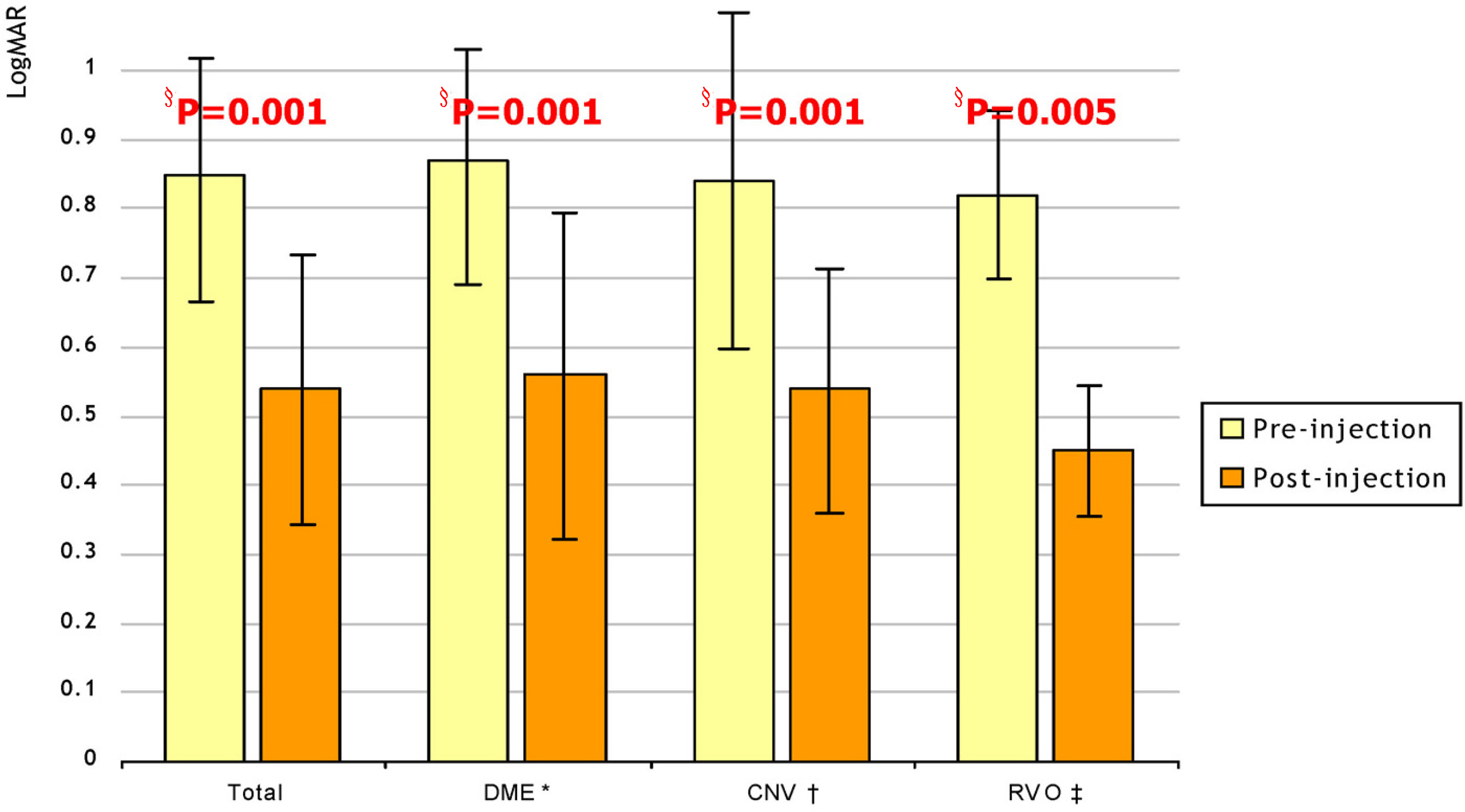
Figure 2. Changes in visual acuity (logMAR) after intravitreal bevacizumab injection (IVB) among diseases. * DME=diabetic macular edema; † CNV=choroidal neovascularization; ‡ RVO=retinal vein occlusion; § P=paired t –test.

Figure 3. Changes in parameters of multifocal electroretinogram before and after intravitreal injection. * r=pearson's correalation.

Figure 4. Changes in central macular thickness after intravitreal bevacizumab injection among diseases.* DME=diabetic macular edema; † CNV=choroidal neovascularization; ‡ RVO=retinal vein occlusion; § p=paired t-test.
Reference
-
References
1. Leung D, Cachianes G, Kuang W, et al. Vascular endothelial growth factor is a secreted angiogenic mitogen. Science. 1989; 246:1306–9.
Article2. Plate KH, Breier G, Weich HA, Risau W. Vascular endothelial growth factor is a potential tumour angiogenesis factor in human gliomas in vivo. Nature. 1992; 359:845–8.
Article3. Rosenfeld PJ, Moshfeghi AA, Puliafito CA. Optical coherence tomography findings after an intravitreal injection of bevacizumab (avastin) for neovascular age-related macular degeneration. Ophthalmic Surg Lasers Imaging. 2005; 36:331–5.
Article4. Seo JW, Park IW. Intravitreal bevacizumab for treatment of diabetic macular edema. Korean J Ophthalmol. 2009; 23:17–22.
Article5. Velez-Montoya R, Fromow-Guerra J, Burgos O, et al. The effect of unilateral intravitreal bevacizumab (avastin), in the treatment of diffuse bilateral diabetic macular edema: a pilot study. Retina. 2009; 29:20–6.6. Chung EJ, Hong YT, Lee SC, et al. Prognostic factors for visual outcome after intravitreal bevacizumab for macular edema due to branch retinal vein occlusion. Graefes Arch Clin Exp Ophthalmol. 2008; 246:1241–7.
Article7. Sutter E, Tran D. The field topography of ERG components in man-1. The photopic luminance response. Vision Res. 1992; 32:433–46.8. Boulton M, Foreman D, Williams G, et al. VEGF localisation in diabetic retinopathy. Br J Ophthalmol. 1998; 82:561–8.
Article9. Adamis A, Miller J, Bernal M, et al. Increase vascularendothelia growth factor levels in the vitreous of eyes withproliferative diabetic retinopathy. Am J Ophthalmol. 1994; 118:445–50.10. Funatsu H, Yamashita H, Ikeda T, et al. Vitreous levels of interleukin-6 and vascular endothelial growth factor are related to diabetic macular edema. Ophthalmology. 2003; 110:1690–6.
Article11. Funatsu H, Yamashita H, Sakata K, et al. Vitreous levels of vascular endothelial growth factor and intercellular adhesion molecule 1 are related to diabetic macular edema. Ophthalmology. 2005; 112:806–16.
Article12. Barakat MR, Kaiser PK. VEGF inhibitors for the treatment of neovascular age-related macular degeneration. Expert Opin Investig Drugs. 2009; 18:637–46.
Article13. Kim JY, Kweon EY, Lee DW, Cho NC. Results of intravitreal bevacizumab for macular edema with retinal vein occlusion and diabetic macular edema. J Korean Ophthalmol Soc. 2008; 49:1275–82.
Article14. Chang MW, KIM SW, Oh IK, et al. Intravitreal triamcinolone injection with or without bevacizumab for diabetic macular edema. J Korean Ophthalmol Soc. 2008; 49:1269–74.
Article15. Lee MH, An JH, Lee JE, Oum BS. Short-term efficacy of intravitreal bavacizumab for polypoidal choroidal vasculopathy. J Korean Ophthalmol Soc. 2009; 50:51–60.
Article16. Lee SW, Kim MS, Kim ES, et al. Long-term results of intravitreal bevacizumab injection for macular edema: retinal vein obstruction and diabetic retinopathy. J Korean Ophthalmol Soc. 2009; 50:211–8.
Article17. Oh SB, Cho WB, Moon JW, Kim HC. Effects and prognostic factors of intravitreal bevacizumab injection on choroidal neovascularization from age-related macular degeneration. J Korean Ophthalmol Soc. 2009; 50:202–10.
Article18. Liu YC, Yang CS, Shyong MP, et al. Intravitreal injection of bevacizumab for the treatment of choroidal neovascularization in a patient with angioid streaks. J Chin Med Assoc. 2009; 72:98–102.
Article19. Arevalo JF, Fromow-Guerra J, Quiroz M, et al. Pan-American Collaborative Retina Study Group.Primary intravitreal bevacizumab (Avastin) for diabetic macular edema: results from the Pan-American Collaborative Retina Study Group at 6-month follow-up. Ophthalmology. 2007; 114:743–50.20. Lee YD, Bae SR. Normal values of positive wave in the multifocal electroretinography in Korean. J Korean Ophthalmol Soc. 2003; 44:850–6.21. Moschos MM, Brouzas D, Apostolopoulos M, et al. Intravitreal use of bevacizumab (Avastin) for choroidal neovascularization due to ARMD: a preliminary multifocal-ERG and OCT study. Multifocal-ERG after use of bevacizumab in ARMD. Doc Ophthalmol. 2007; 114:37–44.22. Shetty R, Pai S, Vincent A, et al. Electrophysiological and structural assessment of the central retina following intravitreal injection of bevacizumab for treatment of macular edema. Doc Ophthalmol. 2008; 116:129–35.
Article
- Full Text Links
-
- Actions
-
Cited
- CITED
-
- Close
- Share
-
- Similar articles
-
- Effect of High-dose Intravitreal Bevacizumab Injection on Refractory Idiopathic Choroidal Neovasculariz
- Multifocal Electroretinogram Findings after Intravitreal Bevacizumab Injection in Choroidal Neovascularization of Age-Related Macular Degeneration
- Long-term Therapeutic Effect of Intravitreal Bevacizumab (Avastin) on Myopic Choroidal Neovascularization
- Intravitreal Bevacizumab Injection for Choroidal Neovascularization Secondary to Laser Photocoagulation for Central Serous Chorioretinopathy
- Choroidal Neovascularization in a Patient with Best Disease
