Two Female Siblings With Bietti Crystalline Retinopathy Without Corneal Dystrophy
- Affiliations
-
- 1Department of Ophthalmology, Seoul National University College of Medicine, Seoul, Korea. hgonyu@snu.ac.kr
- 2Sensory Organ Institute, Medical Research Center, Seoul National University, Seoul, Korea.
- KMID: 2212505
- DOI: http://doi.org/10.3341/jkos.2009.50.7.1120
Abstract
- PURPOSE
To report clinical and functional results in two female siblings with Bietti crystalline retinopathy. CASE SUMMARY: Recently, a 48-year-old female with bilateral intraretinal depositions presented with a complaint of decreased visual acuity and night blindness in both eyes. Several tiny glistening yellow intraretinal crystalline depositions were observed. Fluorescein angiography showed a well-demarcated choriocapillaris filling defect and pigment epithelial window defect. Electrophysiologic tests showed decreased amplitude and OCT scans showed fine intraretinal lesions with increased signal intensity. In addition, a 50-year-old female sibling presented with bilateral yellow, intraretinal crystalline depositions. A choriocapillaris filling defect and pigment epithelial window defect in a fluorescein angiography was observed. Electrophysiologic tests showed severely decreased amplitude. CONCLUSIONS: Two female siblings with Bietti crystalline retinopathy are reported.
Keyword
MeSH Terms
Figure
-

Figure 1. Case 1. (A) Numerous retinal crystals are found throughout the posterior pole and mid-periphery. Pigmentary clumps, atrophy of the retinal pigment epithelium, and choriocapillaris atrophy are also found. (B) An early phase of fluorescein angiography. It shows well defined choriocapillaris filling defects and pigment epithelial window defect. The crystalline deposits do not stain, but some of them block the fluorescence. (C) Indocyanine green angiography shows patchy atrophy of the choriocapillaris and pigment epithelial window defect. (D) OCT scans show intraretinal hyperintense lesions.

Figure 2. Full-field ERG shows reduced amplitude. (A) Case 1, (B) Case 2.
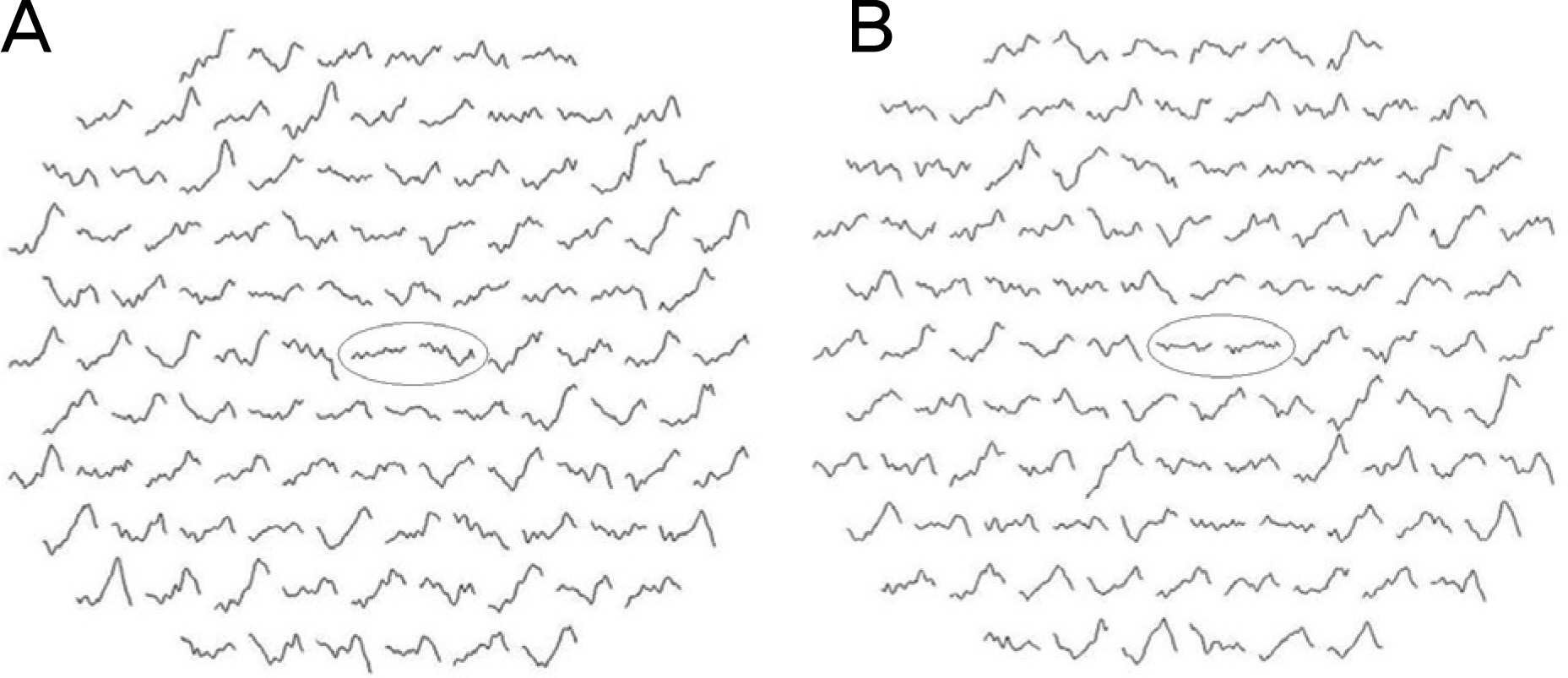
Figure 3. Case 1. Multifocal ERG shows reduced amplitude at the central macular area. (A) right eye, (B) left eye.
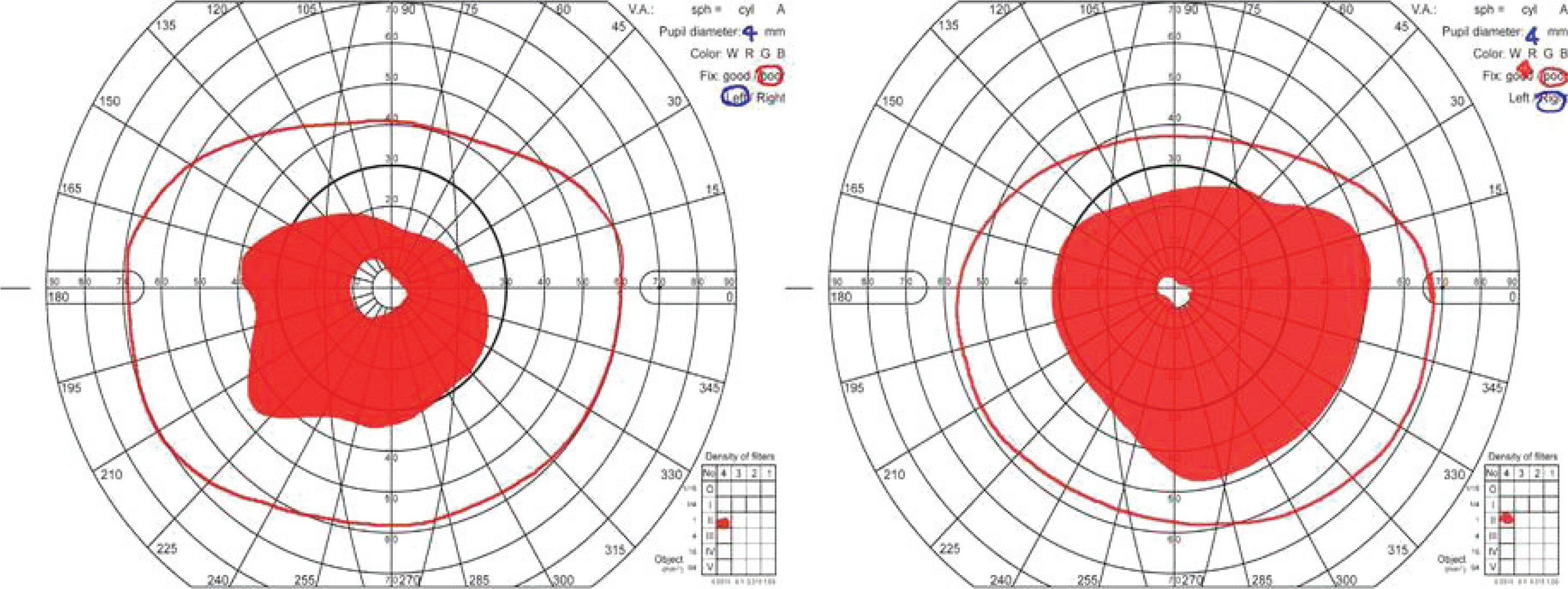
Figure 4. Case 1. Goldmann visual field test shows constriction of the visual field in both eyes.

Figure 5. Case 2. (A) Numerous retinal crystals are found throughout the posterior pole and mid-periphery. Pigmentary clumps, atrophy of the retinal pigment epithelium, and choriocapillaris atrophy are also found. (B) An early phase of fluorescein angiography. It shows generalized choriocapillaris filling defects and pigment epithelial window defect. The crystal line deposits do not stain, but some of them block the fluorescence.

Figure 6. Case 2. (A) Goldmann visual field test shows some peripheral visual field and central visual field remained in the left eye and some peripheral field remained in the right eye. (B) After two years, Goldmann visual field test shows more visual field loss than on the previous test.
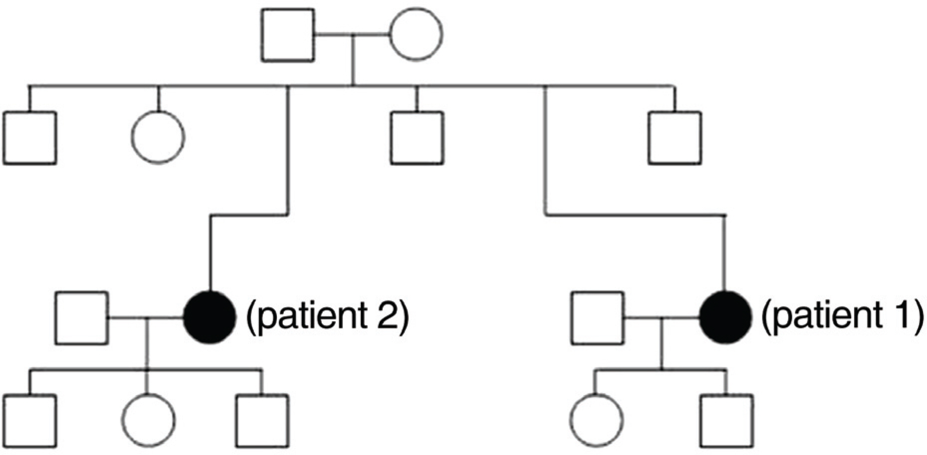
Figure 7. Pedigree chart of the family.
Reference
-
References
1. Bietti GB. Ueber familiares Vorkommen von “Retinitis punctata albescens” (verbunden mit “Dystrophia marginaliscristallinea cornea”): Glitzern des Glaskörpers und anderen degenerativen Augenveränderungen. Klin Monatsbl Augenheilkd. 1937; 99:737–56.2. Bagolini B, Ioli-Spada G. Bietti's tapetoretinal degeneration with marginal corneal dystrophy. Am J Ophthalmol. 1968; 65:53–60.3. Welch RB. Bietti's tapetoretinal degeneration with marginal corneal dystrophy crystalline retinopathy. Trans Am Ophthalmol Soc. 1977; 75:164–79.4. Grizzard WS, Deutman AF, Nijhuis F, de Kerk AA. Crystalline retinopathy. Am J Ophthalmol. 1978; 86:81–8.5. Kaiser-Kupfer MI, Chan CC, Markello TC, et al. Clinical biochemical and pathologic correlations in Bietti's crystalline dystrophy. Am J Ophthalmol. 1994; 118:569–82.
Article6. Hu DN. Ophthalmic genetics in China. Ophthalmic Paediatr Genet. 1983; 2:39–45.
Article7. Lin J, Nishiguchi KM, Nakamura M, et al. Recessive mutations in the CYP4V2 gene in East Asian and Middle Eastern patients with Bietti crystalline corneoretinal dystrophy. J Med Genet. 2005; 42:e38.
Article8. Chan WM, Pang CP, Leung AT, et al. Bietti crystalline retinopathy affecting all 3 male siblings in a family. Arch Ophthalmol. 2000; 118:129–31.9. Bernauer W, Daicker B. Bietti's corneal retinal dystrophy. A 16-year progression. Retina. 1992; 12:18–20.10. Mauldin WM, O'Conner PS. Crystalline retinopathy (Bietti's tapetoretinal degeneration without marginal corneal dystrophy). Am J Ophthalmol. 1981; 92:640–6.
Article11. Richards BW, Brodstein DE, Nussbaum JJ, et al. Autosomal dominant crystalline dystrophy. Ophthalmology. 1991; 98:658–65.
Article12. Miyauchi O, Murayama K, Adachi-Usami E. A family with crystalline retinopathy demonstrating an autosomal dominant inheritance pattern. Retina. 1999; 19:573–4.
Article13. Hahn DK, Park YH. A case of crystalline retinopathy. J Korean Ophthalmol Soc. 1995; 36:142–6.14. Cho HK, Ko SM. A case of crystalline retinopathy. J Korean Ophthalmol Soc. 1997; 38:1628–31.15. Kim HD, Seo MS. Crystalline retinopathy without corneal dystrophy. J Korean Ophthalmol Soc. 2000; 41:1445–50.16. Li A, Jiao X, Munier FL, et al. Bietti crystalline corneoretinal dystrophy is caused by mutations in the novel gene CYP4V2. Am J Hum Genet. 2004; 74:817–26.
Article17. Wada Y, Itabash T, Sato H, et al. Screening for mutations in CYP4V2 gene in Japanese patients with Bietti's crystalline corneoretinal dystrophy. Am J Ophthalmol. 2005; 139:894–9.
Article18. Lee KY, Koh AH, Aung T, et al. Characterization of Bietti crystalline dystrophy patients with CYP4V2 mutations. Invest Ophthalmol Vis Sci. 2005; 46:3812–6.19. Shan M, Dong B, Zhao X, et al. Novel mutations in the CYP4V2 gene associated with Bietti crystalline corneoretinal dystrophy. Mol Vis. 2005; 11:738–43.20. Gekka T, Hayashi T, Takeuchi T, et al. CYP4V2 mutations in two Japanese patients with Bietti's crystalline dystrophy. Ophthalmic Res. 2005; 37:262–9.21. Yoshida A, Nara Y, Takahashi M. Crystalline retinopathy: evaluation of blood-retinal barrier by vitreous fluorophotometry. Jpn J Ophthalmol. 1980; 29:290–300.22. Yagasaki K, Miyake Y. Crystalline retinopathy. Nippon Ganka Gakkai Zasshi. 1986; 90:711–9.23. Takikawa C, Miyake Y, Yagasaki K. Reevaluation of crystalline retinopathy based on corneal findings. Folia Ophthalmol Jpn. 1992; 43:969–78.24. Usui T, Tanimoto N, Takagi M, et al. Rod and cone a-waves in three cases of Bietti crystalline chorioretinal dystrophy. Am J Ophthalmol. 2001; 132:395–402.
Article25. Kretschmann U, Usui T, Ruether K, Zrenner E. Electroretinographic campimetry in a patient with crystalline retinopathy. Ger J Ophthalmol. 1996; 5:399–403.26. Chen H, Zhang M, Huang S, Wu D. Functional and clinical findings in 3 female siblings with crystalline retinopathy. Doc Ophthalmol. 2008; 116:237–43.
Article27. Lai TY, Ng TK, Tam PO, et al. Genotype-phenotype analysis of Bietti's crystalline dystrophy in patients with CYP4V2 mutations. Invest Ophthalmol Vis Sci. 2007; 48:5212–20.
- Full Text Links
-
- Actions
-
Cited
- CITED
-
- Close
- Share
-
- Similar articles
-
- A Case of Crystalline Retinopathy
- A Case of Spontaneous Regression of Schnyder's Crystalline Corneal Dystrophy
- Three Cases of Outer Retinal Tubulation in Bietti's Crystalline Dystrophy
- Acetazolamide for Cystoid Macular Oedema in Bietti Crystalline Retinal Dystrophy
- Crystalline Retinopathy without Corneal Dystrophy
