J Korean Ophthalmol Soc.
2009 Feb;50(2):189-194. 10.3341/jkos.2009.50.2.189.
Corneal Endothelial Changes After ICL (Implantable Collamer Lens) Insertion
- Affiliations
-
- 1Department of Ophthalmology, The St. Vincent's Hospital, The Catholic University of Korea, Suwon, Korea.
- 2Department of Ophthalmology, The KangNam St. Mary's Hospital, The Catholic University of Korea, Seoul, Korea. mskim@catholic.ac.kr
- KMID: 2212039
- DOI: http://doi.org/10.3341/jkos.2009.50.2.189
Abstract
-
PURPOSE: To evaluate the stability of corneal endothelial cells after ICL (implantable contact lens) implantation.
METHODS
Retrospective review of 50 eyes were subjected to implantation of ICL by one surgeon from December 2003 to April 2008. During the follow-up period, the number of eyes with regular follow-up decreased and remained at 18 eyes at the time of a three-year follow-up. Patients were examined preoperatively and at one, three, and six months and one, two, and three years postoperatively. The main outcome measures were the change in corneal endothelial cell density (CD), coefficient of variation (CV), and hexagonality (HA). This analysis was done in all eyes and especially in the 18 eyes with regular follow-up for three years and 11 eyes with changes in CV and HA corresponding to the normal endothelial wound healing process for three years.
RESULTS
Corneal endothelial CD, CV, and HA were not significantly changed compared with their preoperative values throughout the three-year follow-up period for all eyes. In the early postoperative period, there was a slight decrease in CD, but it increased after the second year of follow-up (range: -3.12%~+2.84%); there was a slight increase in CV, but it decreased after the six-month follow-up (-6.63%~+5.56%); and there was a slight decrease in HA, but it increased after the three-month follow-up (range: -0.88%~10.64%). Similarly, there were no significant changes in corneal endothelial cell density in the 18 eyes with regular follow-up or the 11 eyes with changes in CV and HA corresponding to normal endothelial wound healing process for three years.
CONCLUSIONS
The results show stability of corneal endothelial cells after ICL implantation.
MeSH Terms
Figure
-
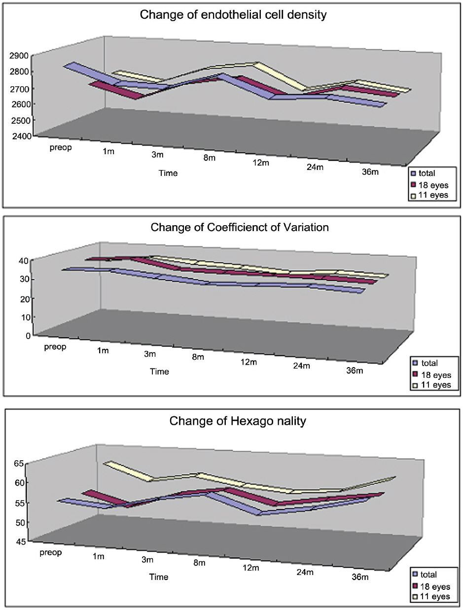
Figure 1. Postoperative change of endothelial cell density, coefficient of variation and hexagonality in total eyes (total), 18 eyes with 3 year follow up (18 eyes), 18 eyes with changes of CV and HA corresponding to the normal wound healing process (11 eyes). There were no significant changes in cell density, coefficient of variation and hexagonality compared with preoperative value.
Reference
-
References
1. Dejaco-Ruhswurm I, Scholz U, Pieh S, et al. Long term endothelial changes in phakic eyes with posterior chamber intraocular lenses. J Cataract Refract Surg. 2002; 28:1589–93.2. Han SY, Lee KH. Long term effect of ICL implantation to treat high myopia. J Korean Ophthalmol Soc. 2007; 48:465–72.3. Chun YS, Lee JH, Lee JM, et al. Outcomes after implantable contact lens for moderater to high myopia. J Korean Ophthalmol Soc. 2004; 45:480–9.4. Lee SY, Cheon HJ, Baek TM, et al. Implantable contact lens to correct high myopia (clinical study with 24 months follow-up). J Korean Ophthalmol Soc. 2000; 41:1515–22.5. Jiménez-Alfaro I, Benítez del Castillo JM, García-Feijoó J, et al. Safety of posterior chamber phakic intraocular lenses for the correction of high myopia. Ophthalmology. 2001; 108:90–9.
Article6. Kim KS, Park SY, Oh JS. Morphometric analysis of the corneal endothelial cells in normal Korean. J Korean Ophthalmol Soc. 1992; 33:320–5.7. Bernard EM, Henry FE, Michael JL. Review of corneal endothelial specular microscopy for FDA clinical trials of refractive procedures, surgical devices, and intraocular drugs and solutions. Cornea. 2008; 27:1–16.8. Bourne WM, Nelson LR, Buller CR, et al. Long term observation of morphologic and functional features of cat corneal endothelium after wounding. Invest Ophthalmol Vis Sci. 1994; 35:891–9.9. Ling TL, Vannas A, Holden BA. Long term changes in corneal endothelial morphology following wounding in the cat. Invest Ophthalmol Vis Sci. 1998; 29:1407–12.10. Ichijima H, Petroll WM, Jester JV, et al. In vivo confocal microscopic studies of endothelial wound healing in rabbit cornea. Cornea. 1993; 12:369–78.
Article11. Odenthal MT, Gan IM, Oosting J, et al. Long term changes in corneal endothelial morphology after discontinuation of low gas permeable contact lens wear. Cornea. 2005; 24:32–8.12. Huang PT, Nelson LR, Bourne WM. The morphology and function of healing cat corneal endothelium. Invest Ophthalmol Vis Sci. 1989; 30:1794–801.13. Pesando PM, Ghiringhello MP, Di Meglio G, Fanton G. Posterior chamber phakic intraocular lens (ICL) for hyperopia: Ten-year follow up. J Cataract Refract Surg. 2007; 33:1579–84.14. Bechmann M, Ullrich S, Thiel MJ, et al. Imaging of posterior chamber phakic intraocular lens by optical coherence tomography. J Cataract Refract Surg. 2002; 28:360–3.
Article15. García-Feijoó J, Alfaro IJ, Cuiña-Sardiña R, et al. Ultrasound Biomicroscopy examination of posterior chamber phakic intraocular lens position. Ophthalmology. 2003; 110:163–72.
Article16. Lackner B, Pieh S, Schmidinger G, et al. Long term results of implantation of phakic posterior chamber intraocular lenses. J Cataract Refract Surg. 2004; 30:2269–76.17. Sanders DR, Doney K, Poco M. ICL in Treatment of Myopia Study Group. United States Food and Drug administration Clinical Trial of the Implantable Collamer Lens (ICL) for moderate to high myopia. Ophthalmology. 2004; 111:1683–92.18. Edelhauser HF, Sanders DR, Azar R, Lamielle H. ICL in Treatment of Myopia Study Group. Corneal endothelial assessment after ICL implantation. J Cataract Refract Surg. 2004; 30:576–83.19. Bourne WM, Nelson LR, Hodge DO. Central corneal endothelial cell changes over a ten-year period. Invest Ophthalmol Vis Sci. 1997; 38:779–82.20. Budo C, Hesslooehl JC, Izak M, et al. Multicenter study of the Artisan phakic intraocular lens. J Cataract Refract Surg. 2000; 26:1163–71.
Article21. Chang SW, Hu FR, Lin LL. Effects of contact lenses on corneal endothelium-a morphological and functional study. Ophthalmologica. 2001; 215:197–203.22. Sibug ME, Datiles MB, Kashima K, et al. Specular micro-scopiy studies on the corneal endothelium after cessation of contact lens wear. Cornea. 1991; 10:395–401.
- Full Text Links
-
- Actions
-
Cited
- CITED
-
- Close
- Share
-
- Similar articles
-
- Long-term Clinical Outcomes of Implantable Collamer Lens
- Effects of a Novel Push-through Technique Using the Implantable Collamer Lens Injector System for Graft Delivery during Endothelial Keratoplasty
- The Analysis of Vault Change after Posterior Chamber Phakic Intraocular Lens Size Exchange
- Axial Length Change after Implantable Collamer Lens Implantation
- Partial Visual Rehabilitation Using a Toric Implantable Collamer Lens in a Patient with Keratoconus: A Case Report with 20 Months of Follow-up
