Adenosquamous Carcinoma Arising in Congenital Choledochal Cyst
- Affiliations
-
- 1Department of Surgery, Seoul National University Boramae Hospital, Seoul, Korea. ahnyj@brm.co.kr
- 2Department of Pathology, Seoul National University Hospital, Seoul, Korea.
- KMID: 2211960
- DOI: http://doi.org/10.4174/jkss.2010.78.5.325
Abstract
- Adenosquamous carcinoma arising in congenital choledochal cyst is very rare and herein we report a case thereof. A 37-year-old woman was referred for further evaluation of pancreas head mass and a hepatic nodule on CT. She had been diagnosed with congenital choledochal cyst at 22-years-old and received Roux-en-Y choledochojejunostomy at that time. Endoscopic ultrasonography-guided biopsy proved the pancreas head mass as a squamous cell carcinoma and liver biopsy also proved the liver mass as a metastatic squamous cell carcinoma. We performed pancreaticoduodenectomy and tumorectomy of metastatic liver nodule. Grossly, the primary lesion was located at intrapancreatic portion of choledochal cyst. Histologically, the primary lesion and hepatic nodule was metastatic adenosquamous carcinoma. So far, there have been only three cases of adenosquamous carcinoma arising in congenital choledochal cyst reported in English-language literature. This is another case and the first case reported in Korea.
Keyword
MeSH Terms
Figure
-
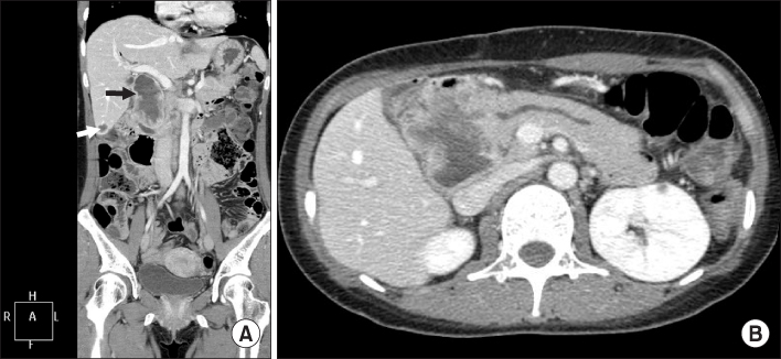
Fig. 1 (A) 3D abdominal CT shows 6 cm sized heterogeneous enhancing mass (black arrow) with internal necrotic portion between pancreas and duodenum and a 1.2 cm sized hepatic nodule (white arrow) at segment 6. (B) 2D abdominal CT shows pancreas head mass with duct dilatation.

Fig. 2 Gross specimen. (A) Gross photography of the specimen shows dilated distal CBD and encircling mass within it. (B) Transverse section of the mass shows its solid and heterogenous characteristic.
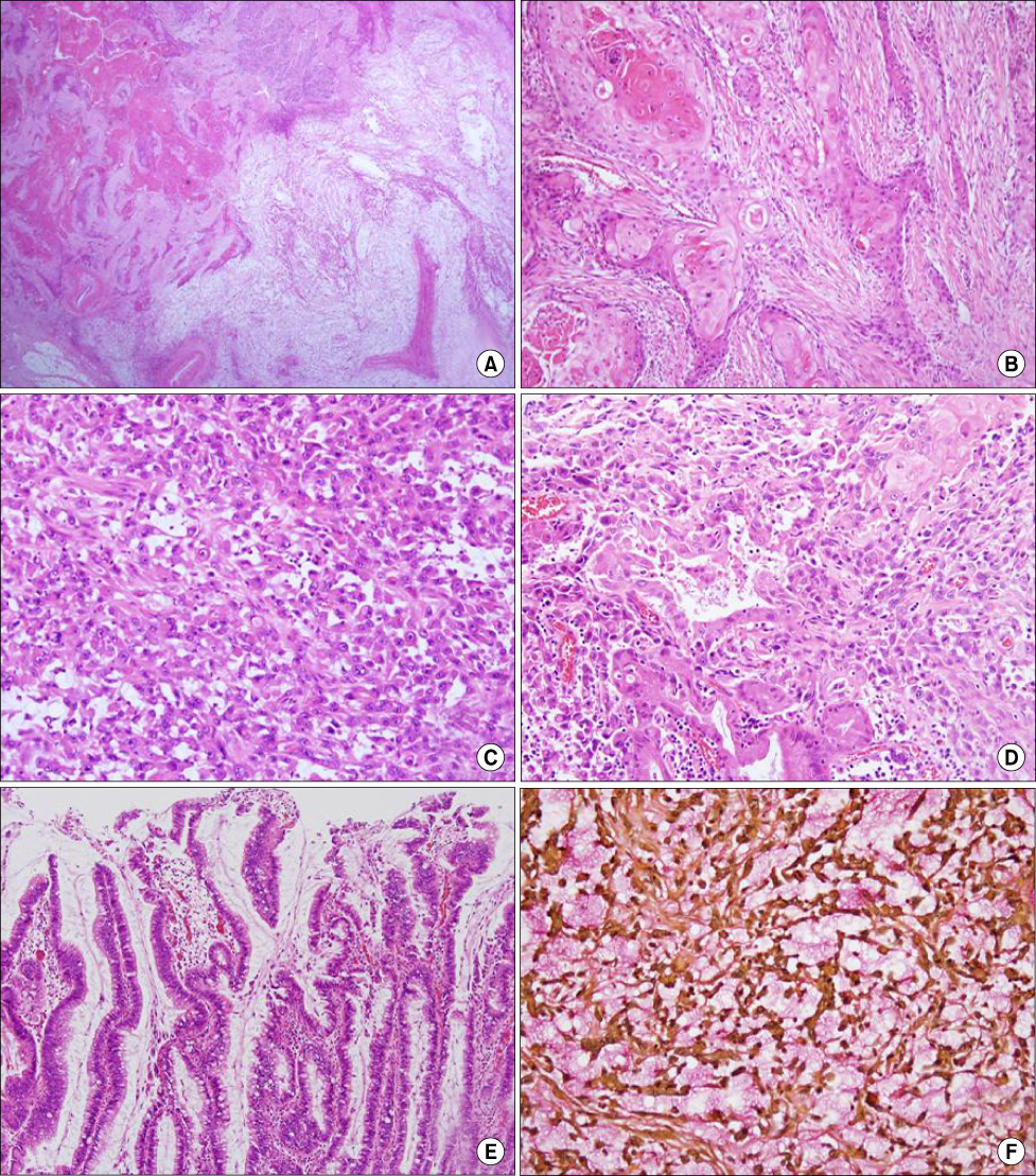
Fig. 3 Microscopic findings of the tumor - (A) The tumor was composed of squamous cell carcinoma component (left side) and adenocarcinoma component (right side) (H&E stain, ×10). (B) Squamous cell carcinoma component: well differentiated squamous cell carcinoma forming keratin pearl (H&E stain, ×100). (C) Adenocarcinoma component: sheet of atypical cell with mucin production (H&E stain, ×200). (D) Relatively well-formed glands in transition zone between squamous cell carcinoma and adenocarcinoma component (H&E stain, ×200). (E) High grade dysplasia in adjacent bile duct epithelium (H&E stain, ×100). (F) Mucicarmine was positive in mucin pool of adenocarcinoma component (mucicarmine stain, ×200).
Reference
-
1. Solcia E, Capella C, Kloppel G. . Tumors of the Pancreas. 1997. Washington, D.C.: Armed Forces Institute of Pathology;88–91. (Atlas of tumot pathology. Third series, fasc. 20).2. Cao SH. Extrahepatic bile duct cancer. Report of 106 cases. Chin Med J (Engl). 1992. 105:622–629.3. Okamura K, Hayakawa H, Kuze M, Takahashi H, Kosaka A, Mizumoto R, et al. Triple carcinomas of the biliary tract associated with congenital choledochal dilatation and pancreaticobiliary maljunction. J Gastroenterol. 2000. 35:465–471.4. Terada T. Adenosquamous carcinoma in a congenital choledochal cyst associated with pancreatico-biliary maljunction. Pathol Int. 2009. 59:482–485.5. Terada T, Nakanuma Y. Congenital biliary dilatation in autosomal dominant adult polycystic disease of the liver and kidneys. Arch Pathol Lab Med. 1988. 112:1113–1116.6. Kamisawa T, Okamoto A, Tsuruta K, Tu Y, Egawa N. Carcinoma arising in congenital choledochal cysts. Hepatogastroenterology. 2008. 55:329–332.7. Fieber SS, Nance FC. Choledochal cyst and neoplasm: a comprehensive review of 106 cases and presentation of two original cases. Am Surg. 1997. 63:982–987.8. Price L, Kozarek R, Agoff N. Squamous cell carcinoma arising within a choledochal cyst. Dig Dis Sci. 2008. 53:2822–2825.9. Nonomura A, Mizukami Y, Matsubara F, Ueda H. A case of choledochal cyst associated with adenocarcinoma exhibiting sarcomatous features. J Gastroenterol. 1994. 29:669–675.10. Lantsberg L, Khodadadi J, Goldstein J. Adenosquamous carcinoma of the common bile duct: a case report. J Surg Oncol. 1986. 33:140–142.
