Inflammatory Pseudotumor of Splenic Hilar Node Mimicking Gastric Submucosal Tumor
- Affiliations
-
- 1Department of Surgery, Yeungnam University College of Medicine, Daegu, Korea. sksong@med.yu.ac.kr
- KMID: 2211950
- DOI: http://doi.org/10.4174/jkss.2010.78.3.199
Abstract
- Inflammatory pseudotumor (IPT) is an uncommon clinical condition characterized by proliferation of spindle cells, inflammatory cells, and small vessels. IPT has been reported in various anatomical sites, including the orbit, lung, liver, spleen, and so on. IPT of the lymph node is very rare. We recently experienced a 65-year-old woman diagnosed with IPT of the lymph node in the splenic hilar, or distal supra-pancreatic area, mimicking gastric submucosal tumor. The tumor was removed without event using the laparoscopic method. This lesion in the splenic hilum is extremely rare and has not been cited in the current literature. We describe such a rare case of IPT with a review of the literature.
Figure
-

Fig. 1 (A) Gastroscopic finding showed bulging mucosa in the greater curvature side of the gastric body. (B) Endoscopic ultrasound finding showed a hypoechoic lesion in the muscularis propria layer, 2.5×2.0 cm in diameter, in the greater curvature side of the gastric body.
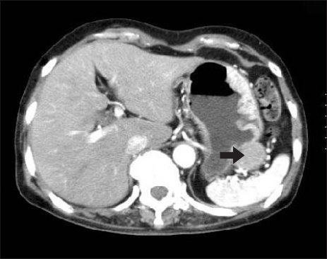
Fig. 2 Abdominal CT finding. The black arrow indicates a well-defined exophytic mass, with early enhancing nature in the greater curvature side of the gastric body.
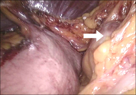
Fig. 3 Intraoperative finding. The white arrow indicates a well-defined mass in a location between the upper border of the distal pancreas and the splenic hilum.
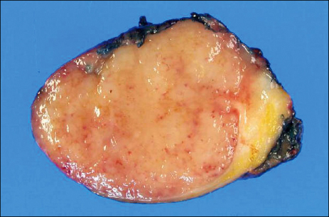
Fig. 4 The lymph node in the specimen measured 3.3×3.0×2.2 cm and weighed 12.7 g.

Fig. 5 Histopathologic finding. (A) This histopathologic features were consistent with lymph node (H&E, ×10). (B) The lymph node showed plasma cell infiltration and sclerosis in the focal area. The inflammatory cells infiltrate, including plasma cells, lymphocytes, eosinophils, and sclerosis were also noted in the perinodal soft tissues (H&E, ×100).
Reference
-
1. Umiker WO, Iverson L. Postinflammatory tumors of the lung; report of four cases simulating xanthoma, fibroma, or plasma cell tumor. J Thorac Surg. 1954. 28:55–63.2. Facchetti F, De Wolf Peeters C, De Wever I, Frizzera G. Inflammatory pseudotumor of lymph nodes. Immunohistochemical evidence for its fibrohistiocytic nature. Am J Pathol. 1990. 137:281–289.3. Perrone T, De Wolf-Peeters C, Frizzera G. Inflammatory pseudotumor of lymph nodes. A distinctive pattern of nodal reaction. Am J Surg Pathol. 1988. 12:351–361.4. Knockaert DC, Schuermans A, Vlayen J, Dewolf-Peeters C, Bobbaers HJ. Fever of unknown origin due to inflammatory pseudotumour of lymph nodes. Acta Clin Belg. 1998. 53:367–370.5. Miras-Parra FJ, Parra-Ruiz J, Gomez-Morales M, Gomez-Jimenez FJ, de la Higuera-Torres-Puchol J. Inflammatory pseudotumor of lymph nodes with focal infiltration in liver and spleen. Dig Dis Sci. 2003. 48:2003–2004.6. Gunny RS, Akhbar N, Connor SE. CT and MRI appearances of inflammatory pseudotumour of the cervical lymph nodes. Br J Radiol. 2005. 78:651–654.7. Kemper CA, Davis RE, Deresinski SC, Dorfmann RF. Inflammatory pseudotumor of intra-abdominal lymph nodes manifesting as recurrent fever of unknown origin: a case report. Am J Med. 1991. 90:519–523.8. Kojima M, Nakamura S, Shimizu K, Hosomura Y, Ohno Y, Itoh H, et al. Inflammatory pseudotumor of lymph nodes: clinicopathologic and immunohistological study of 11 Japanese cases. Int J Surg Pathol. 2001. 9:207–214.9. Fisher RG, Wright PF, Johnson JE. Inflammatory pseudotumor presenting as fever of unknown origin. Clin Infect Dis. 1995. 21:1492–1494.10. Andrade DM, Martins SJ, Paz O, Cardozo JB, Novaes AE, Santiago MB. Inflammatory pseudotumor: A diagnostic dilemma. Eur J Intern Med. 2006. 17:514–516.
- Full Text Links
-
- Actions
-
Cited
- CITED
-
- Close
- Share
-
- Similar articles
-
- Advantages of Splenic Hilar Lymph Node Dissection in Proximal Gastric Cancer Surgery
- Disadvantages of Complete No. 10 Lymph Node Dissection in Gastric Cancer and the Possibility of Spleen-Preserving Dissection: Review
- Subepithelial Lesion of the Gastric Fundus Caused by an Accessory Spleen
- A Case of Splenic Artery Aneurysm Mimicking Gastric Submucosal Tumor
- Gastric Pseudotumoral Lesion Caused by a Fish Bone Mimicking a Gastric Submucosal Tumor
