Clear Cell Adenocarcinoma Arising from Adenofibroma in a Patient with Endometriosis of the Ovary
- Affiliations
-
- 1Department of Pathology, Chosun University School of Medicine, Gwangju, Korea. sclim@chosun.ac.kr
- KMID: 2211383
- DOI: http://doi.org/10.4132/jptm.2015.08.07
Abstract
- Ovarian clear cell adenocarcinomas (CCACs) are frequently associated with endometriosis and, less often with clear cell adenofibromas (CCAFs). We encountered a case of ovarian CCAC arising from benign and borderline adenofibromas of the clear cell and endometrioid types with endometriosis in a 53-year-old woman. Regions of the adenofibromas showed transformation to CCAC and regions of the endometriosis showed atypical endometriotic cysts. This case demonstrates that CCAC can arise from CCAF or endometriosis.
Keyword
MeSH Terms
Figure
-
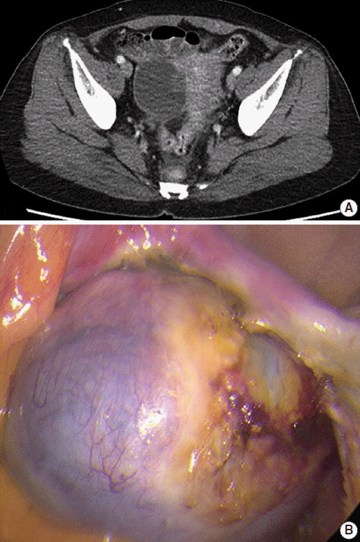
Fig. 1. Abdominal computed tomography (CT) and laparoscopic findings. (A) Abdominal computed tomography showing a 5.5-cm cystic mass in the right ovary. (B) Laparoscopy findings reveal a fluid containing cystic mass with a smooth surface and focal hemorrhage.
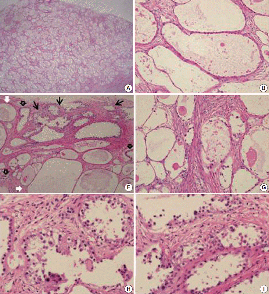
Fig. 2. Histopathologic findings of the solid part of the mass. (A) The compact arrangement of variably-sized tubulocystic structures in the stroma is consistent with adenofibroma. The cell lining consisted of flattened indiscernible cells or flat cuboidal cells (B) and polygonal cells with abundant clear cytoplasm (C). (D, E) In some areas, stratified epithelium shows tiny buds with atypical nuclei. (F) The transitional zone from benign (white arrows) to borderline (black open arrows) clear cell adenofibromas to clear cell adenocarcinoma (black arrows). (G) Higher magnification shows benign (left) and atypical (right) adenofibromas. (H, I) Area of clear cell adenocarcinoma shows a tubulocystic pattern with hobnail, cuboidal, or flat atypical lining cells characterized by nuclear enlargement and hyperchromasia, and foci of altered stromal responses.
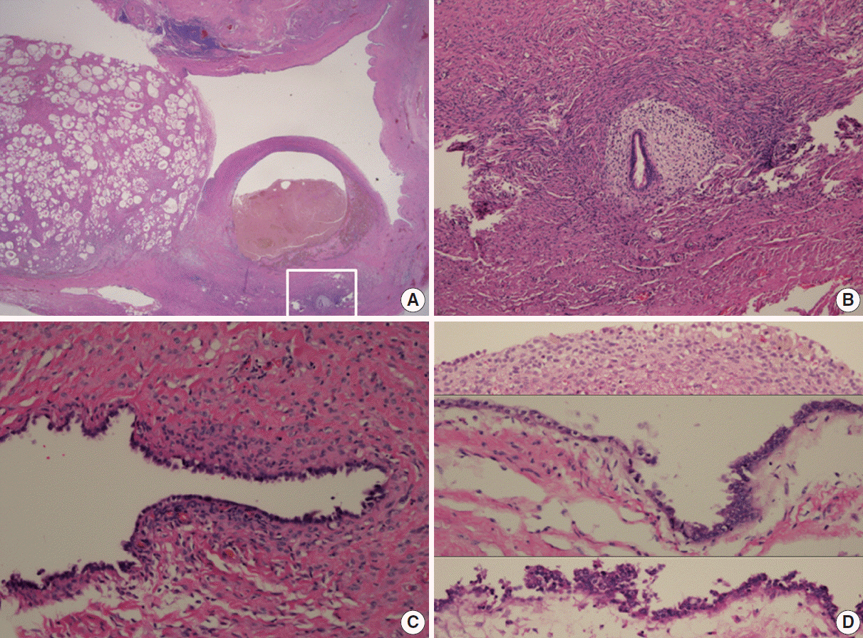
Fig. 3. Histopathologic findings of adenofibroma, endometriotic cyst, and endometriosis (A). Higher magnification of the inset shows endometriosis (B). (C) Endometriotic cyst with tubal-type epithelium overlying scant endometrial-type stroma. (D) Endometriotic cyst with simple cuboidal epithelium overlying endometrial stroma with hemosiderin pigmentation (upper) and gradual transition to nuclear atypia demonstrating stratification with hyperchromatic, enlarged, and irregular nuclei with prominent nucleoli (middle and lower).
Reference
-
1. Seidman JD, Russell P, Kurman RJ. Surface epithelial tumors of the ovary. In : Kurman RJ, editor. Blaustein’s pathology of the female genital tract. 5th ed. New York: Springer-Verlag;2001. p. 791–904.2. Vercellini P, Parazzini F, Bolis G, et al. Endometriosis and ovarian cancer. Am J Obstet Gynecol. 1993; 169:181–2.
Article3. Fukunaga M, Nomura K, Ishikawa E, Ushigome S. Ovarian atypical endometriosis: its close association with malignant epithelial tumours. Histopathology. 1997; 30:249–55.
Article4. Tavassoli FA, Devilee P. World Health Organization classification of tumours of pathology and genetics of tumours of the breast and female genital organs. Lyon: IARC Press;2003. p. 218–28.5. Bell DA, Scully RE. Benign and borderline clear cell adenofibromas of the ovary. Cancer. 1985; 56:2922–31.
Article6. Roth LM, Langley FA, Fox H, Wheeler JE, Czernobilsky B. Ovarian clear cell adenofibromatous tumors: benign, of low malignant potential, and associated with invasive clear cell carcinoma. Cancer. 1984; 53:1156–63.
Article7. Yamamoto S, Tsuda H, Yoshikawa T, et al. Clear cell adenocarcinoma associated with clear cell adenofibromatous components: a subgroup of ovarian clear cell adenocarcinoma with distinct clinicopathologic characteristics. Am J Surg Pathol. 2007; 31:999–1006.
Article8. Yamamoto S, Tsuda H, Takano M, Hase K, Tamai S, Matsubara O. Clear-cell adenofibroma can be a clonal precursor for clear-cell adenocarcinoma of the ovary: a possible alternative ovarian clear-cell carcinogenic pathway. J Pathol. 2008; 216:103–10.
Article9. Jiang X, Morland SJ, Hitchcock A, Thomas EJ, Campbell IG. Allelotyping of endometriosis with adjacent ovarian carcinoma reveals evidence of a common lineage. Cancer Res. 1998; 58:1707–12.10. Obata K, Hoshiai H. Common genetic changes between endometriosis and ovarian cancer. Gynecol Obstet Invest. 2000; 50 Suppl 1:39–43.
Article11. Sato N, Tsunoda H, Nishida M, et al. Loss of heterozygosity on 10q23.3 and mutation of the tumor suppressor gene PTEN in benign endometrial cyst of the ovary: possible sequence progression from benign endometrial cyst to endometrioid carcinoma and clear cell carcinoma of the ovary. Cancer Res. 2000; 60:7052–6.12. Sampson JA. Endometrioid carcinoma of the ovary, arising in endometrial tissue in that organ. Arch Surg. 1925; 10:1–72.13. Russell P. The pathological assessment of ovarian neoplasms. I: Introduction to the common ‘epithelial’ tumours and analysis of benign ‘epithelial’ tumours. Pathology. 1979; 11:5–26.
Article14. Scott RB. Malignant changes in endometriosis. Obstet Gynecol. 1953; 2:283–9.15. Kao GF, Norris HJ. Unusual cystadenofibromas: endometrioid, mucinous, and clear cell types. Obstet Gynecol. 1979; 54:729–36.16. Russell P, Merkur H. Proliferating ovarian “epithelial” tumours: a clinico-pathological analysis of 144 cases. Aust N Z J Obstet Gynaecol. 1979; 19:45–51.
Article17. Bell DA. Ovarian surface epithelial-stromal tumors. Hum Pathol. 1991; 22:750–62.
Article18. Zhao C, Wu LS, Barner R. Pathogenesis of ovarian clear cell adenofibroma, atypical proliferative (borderline) tumor, and carcinoma: clinicopathologic features of tumors with endometriosis or adenofibromatous components support two related pathways of tumor development. J Cancer. 2011; 2:94–106.
Article19. Nishikimi K, Kiyokawa T, Tate S, Iwamoto M, Shozu M. ARID1A expression in ovarian clear cell carcinoma with an adenofibromatous component. Histopathology. 2015; Apr. 23. [Epub]. http://dx.doi.org/10.1111/his.12721.
- Full Text Links
-
- Actions
-
Cited
- CITED
-
- Close
- Share
-
- Similar articles
-
- Clear Cell Adenocarcinoma of the Urinary Bladder Accompanied by Vesical Endometriosis
- Clear cell carcinoma arising in a Cesarean section scar endometriosis: a case report
- Two cases of thromboembolism in clear cell ovarian adenocarcinoma
- A Case Report of Bilateral Squamous Cell Carcinoma Arising in Endometriosis of the Ovary
- A case of clear cell carcinoma arising from the endometriosis of the paraovarian cyst
