A 6-year Observation of Multiple Spinal Schwannomas Before Excision: 1 Case Report
- Affiliations
-
- 1Department of Orthopedic Surgery, Seoul Sacred Heart General Hospital, Seoul, Korea. adkajs@hanmail.net
- KMID: 2209697
- DOI: http://doi.org/10.4184/jkss.2003.10.3.277
Abstract
- Spinal schwannoma is a slow growing symptomatic tumor which is derived from Schwann cells of peripheral nerves. Most reported cases have been single lesion, while multiple schwannomas have invariably documented one manifestation of von Recklinghausen's disease. Nevertheless, we observed a case of independent, multiple, spinal schwannomas for 6 years before excision. We report the growing velocity and progression of neurologic symptoms and neurologic changes before excision and present a literature review.
Keyword
Figure
-

Fig. 1. (A) T1 weighted sagittal MR image which shows intermediate signal double round intradural masses at T12 and L3 level respectively. (B) T2 weighted sagittal MR image which shows no noticeable intradural mass.

Fig. 2. (A) Gd-enhanced sagittal MR image which shows distinctive high signal intradural extramedullary masses at T12 and L3 level respectively and slight olisthesis of L4 body. (B) Gd-enhanced transverse MR image which shows left eccentric intradural extramedullary masses, above T12, below L3.
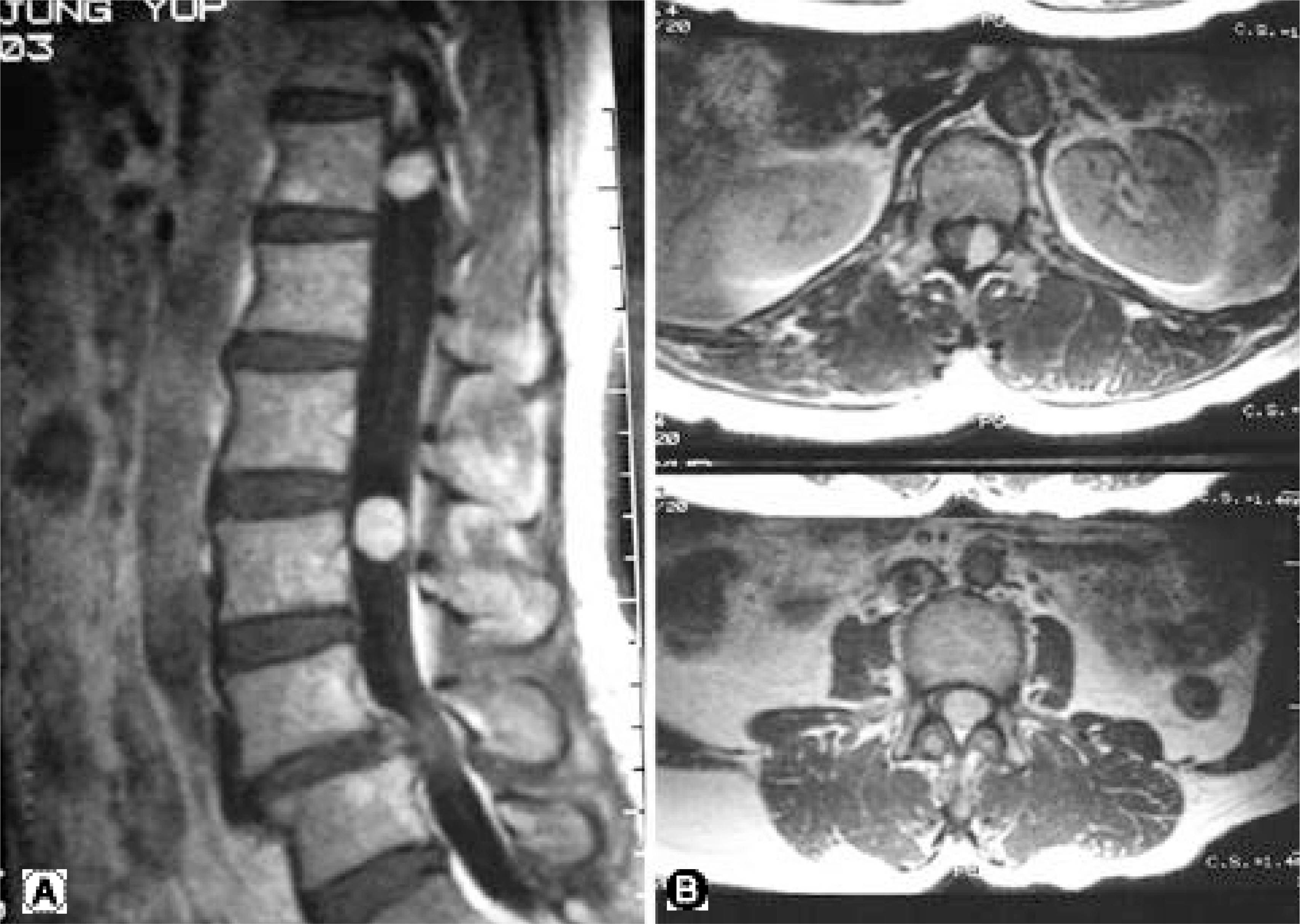
Fig. 3. (A, B) MR image of same patient 3 years later with same facilities and same conditions and it shows more enlarged thoracic and lumbar masses and progression of olisthesis.
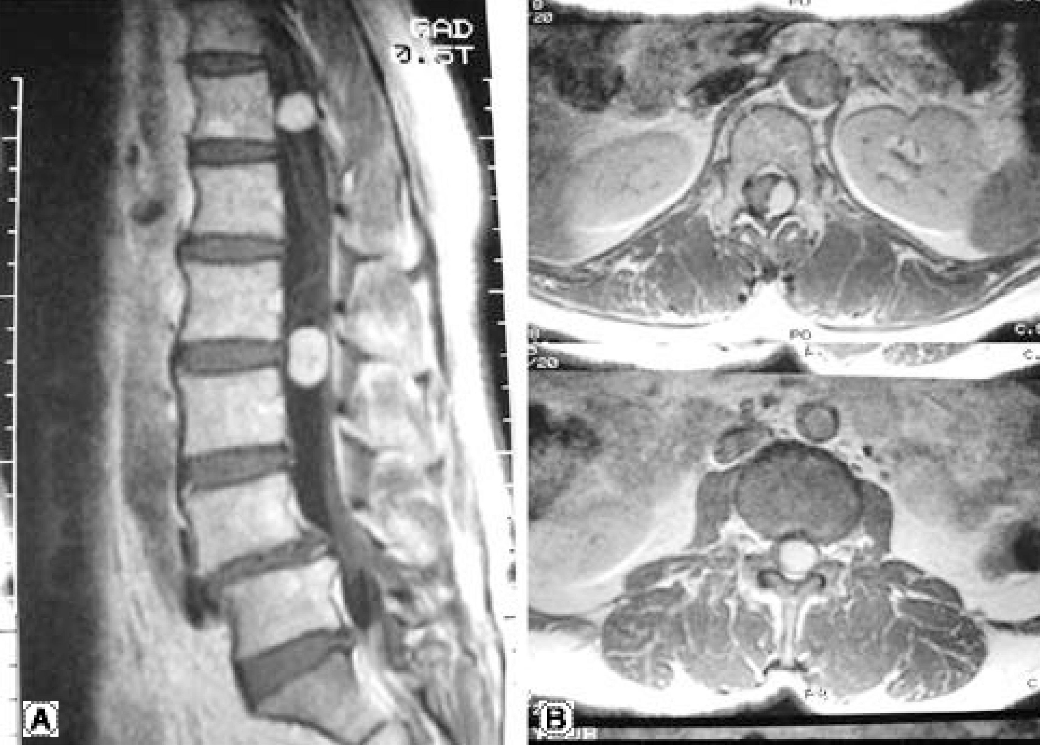
Fig. 4. (A, B) MR image of same patient 6 years later with same facilities and same conditions and it shows more enlarged thoracic and lumbar mass and progression of olisthesis.

Fig. 5. Thoracic mass which containing few intermingled nerve fibers.

Fig. 6. Tumor showing celluar area, Antoni A(arrow) including Verocay bodies(open arrow) as well as looser hypocellular area, Antoni B(arrow head). (H&E stain, × 100)

Fig. 7. Graph which shows growing of tumors according to time.
Cited by 1 articles
-
The Surgical Treatment for Spinal Intradural Extramedullary Tumors
Dong-Ki Ahn, Hoon-Seok Park, Dae-Jung Choi, Kwan-Soo Kim, Tae-Woo Kim, Soon-Youl Park
Clin Orthop Surg. 2009;1(3):165-172. doi: 10.4055/cios.2009.1.3.165.
Reference
-
1). Inoue S, Kataoka H, Tajima N, et al. Assessment of treatment for low back pain. J Jpn Orthop Assoc. 60:391–4. 1986; (cited from Miyakoshi N, Abe E, Shimada Y, Okuyama K, Suzuki T, Sato K: Outcome of One-Level Posterior Lumbar Interbody Fusion for Spondylolisthesis and Postoperative Intervertebral Disc Degeneration Adjacent to the Fusion. Spine 2000;25: 1837-1842).2). Kim P, Ebersold MJ, Onofrio BM, Quast LM. Surgery of spinal nerve schwannoma. Risk of neurological defecit after resection of involved root. J Neurosurg. 1989; 71:810–814.3). Bridwell KH, DeWald RL. The textbook of spinal surgery. 2nd ed.Philadelphia, New York: Lippincott-Raven;p. 2109–2111. 1996.4). Broager B. Spinal neurinoma. A clinical study comprising 44 cases. Acta Psychatr Neurol Scand. 85:1–241. 1953.(cited from Cervoni L, Celli P, Scarpinati M, Cantore G: Neurinomas of the cauda equina. Clinical analysis of 40 surgical cases. Acta Neurochir 1994;127: 199-202).5). Cervoni L, Celli P, Scarpinati M, Cantore G. Neurinomas of the cauda equina. Clinical analysis of 40 surgical cases. Acta Neurochir. 1994; 127:199–202.
Article6). Schneider RC, Kahn EA, Crosby ECC. Spine and spinal cord tumors. McGauley JL, editor. Correlative Neurosurgery. 3rd ed.Springfield: Charles C Thomas;p. 975. 1982.7). Wilkins RH, Rengachary SS. Spinal intradural tumors. Stein BM, editor. Neurosurgery. New York: McGraw-Hill;p. 1048. 1985.
- Full Text Links
-
- Actions
-
Cited
- CITED
-
- Close
- Share
-
- Similar articles
-
- Multicentric Spinal Schwannomas without any Evidence of Neurofibromatosis Type2
- Multiple Schwannomas: Case Report
- Multiple Spinal Tumors with Meningiomas and Schwannomas
- A Case of Multiple Schwannomas of the Trigeminal Nerves, Acoustic Nerves, Lower Cranial Nerves, Brachial Plexuses and Spinal Canal: Schwannomatosis or Neurofibromatosis?
- Three Cases of Primary Spinal Malignant Schwannomas: Case Reports
