Imaging Findings of Malignant Fibrous Histiocytoma of the Breast: A Case Report
- Affiliations
-
- 1Department of Radiology, Kangbuk Samsung Hospital, Sungkyunkwan University School of Medicine, Korea.
- 2Department of Pathology, Kangbuk Samsung Hospital, Sungkyunkwan University School of Medicine, Korea.
- 3Department of General Surgery, Kangbuk Samsung Hospital, Sungkyunkwan University School of Medicine, Korea.
- 4You and Me Surgery, Jeonju, Korea.
- KMID: 2208927
- DOI: http://doi.org/10.3348/jksr.2010.62.3.295
Abstract
- A malignant fibrous histiocytoma (MFH) is the most common soft tissue sarcoma encountered during adulthood, but the breast is not a common site of involvement for MFH. Several investigators have reported the histopathological and biological features of a MFH involving the breast, but only a few reports have focused on the imaging findings of breast MFHs. To emphasize the importance of arriving at a preoperative diagnosis for the treatment implications, we report here the imaging findings, including the mammography, US and MRI findings, for a MFH of the breast of a 53-year-old woman who presented with a rapid growing huge mass in the right breast.
MeSH Terms
Figure
-
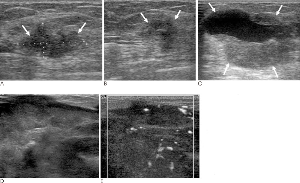
Fig. 1 The ultrasound exam that was done before the VABB. A. There is a 1.87 cm × 1.12 cm sized, irregular, parallel, microlobulated hypoechoic mass in the 10 o'clock direction of the right breast (arrows). B. On the ultrasound exam that was done 3 months after the VABB, there is a 1 cm sized irregular anechoic lesion at the biopsy site (arrows). C. On the ultrasound exam performed 9 months after the VABB, there is a 3.5 × 2.8 × 3 cm sized round, circumscribed mixed echoic mass with posterior enhancement and a partial cystic component at the biopsy site of the right breast (arrows). D. The ultrasound exam performed 4 months afterward reveals a well-defined, huge heterogenous echogenic mass with variable cystic areas that occupy almost the entire right breast. E. The mass shows high vascularity in the solid portion on power Doppler US.
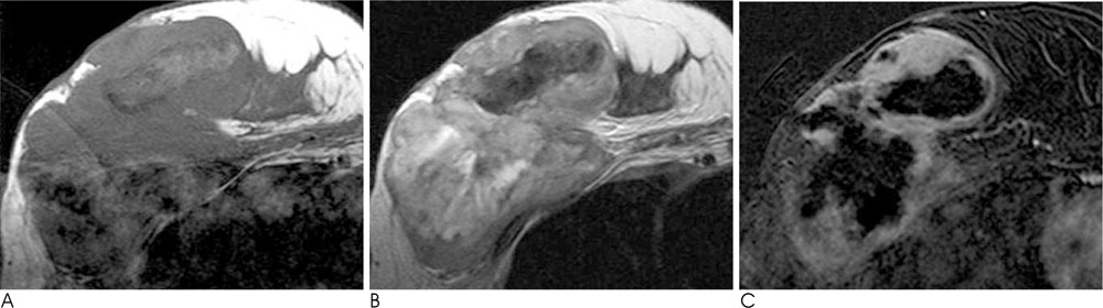
Fig. 2 MR images of the right breast. A, B. There is a 12 cm sized, smoothly well-defined multilobulated intermediate signal intensity mass on the T1 weighted image (A) and the T2 weighted MR image (B). The mass shows central variable, mixed heterogeneous (from very high to very low) signal intensity and peripheral intermediate signal intensity. C. The Gd-enhanced scan shows the early rapid irregular thick peripheral rim enhancement of the mass.
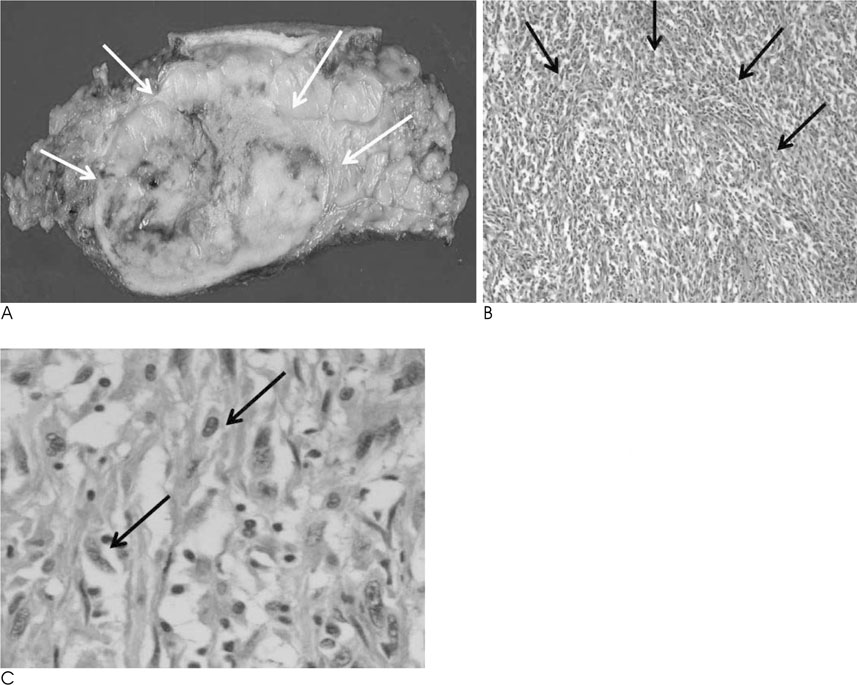
Fig. 3 On the gross specimen, the tumor mass has a relatively well-delineated and lobulating margin, and the mass measures 16×12×6 cm. A. The cut surface is grayish yellow with a "fish flesh" appearance and necrosis and hemorrhage (arrows). B. The histopathology findings of the breast MFH (H & E stain, ×200, ×400) show a diffuse proliferation of atypical spindle cells with a whorling pattern (arrows). C. The tumor cells exhibit round to oval vesicular nuclei with plump eosinophilic or foamy granular cytoplasm (arrows).
Reference
-
1. Murphey MD, Gross TM, Rosenthal HG. From the archives of the AFIP. Musculoskeletal malignant fibrous histiocytoma: radiologicpathologic correlation. Radiographics. 1994; 14:807–826.2. Son E, Park J, Jeon H, Cho S. Malignant fibrous histiocytoma (MFH) in axilla. Yonsei Med J. 2004; 45:736–738.3. Oh SJ, Kim KM, Hong TH, Park WC, Kim JS, Jung SS. Giant cell malignant fibrous histiocytoma of the breast: a case report. J Korean Med Sci. 2004; 19:477–480.4. Hocevar M, Marinsek ZP, Zidar A. Myxofibrosarcoma of the breast as an unusual variant of malignant fibrous histiocytoma: report of a case. Surg Today. 2004; 34:752–754.5. Kijima Y, Umekita Y, Yoshinaka H, Taguchi S, Owaki T, Funasako Y, et al. Stromal sarcoma with features of giant cell malignant fibrous histiocytoma. Breast Cancer. 2007; 14:239–244.6. Ugurlu K, Turgut G, Kabukcuoglu F, Ozcan H, Sanus Z, Bas L. Malignant fibrous histiocytoma developing in a burn scar. Burns. 1999; 25:764–767.7. Ajisaka H, Maeda K, Uchiyama A, Miwa A. Myxoid malignant fibrous histiocytoma of the breast: report of a case. Surg Today. 2002; 32:887–890.
- Full Text Links
-
- Actions
-
Cited
- CITED
-
- Close
- Share
-
- Similar articles
-
- Imaging Findings of Metastatic Breast Malignant Fibrous Histiocytoma: A Case Report
- Post-Radiation Malignant Fibrous Histiocytoma Following Treatment of Breast Cancer: A Case Report with Imaging Findings
- A Case of the retroperitoneal Malignant Fibrous Histiocytoma
- Malignant Fibrous Histiocytoma: A Case Report
- Malignant Fibrous Histiocytoma in the Breast: A Case Report
