Myositis Ossificans of the Psoas Muscle After Compression Fracture of Lumbar Spine: CT and MR Imaging Findings
- Affiliations
-
- 1Department of Radiology, Hallym University College of Medicine, Hangang Sacred Heart Hospital, Korea. lgkhope@nate.com
- 2Department of Radiology, Dongguk University College of Medicine, Gyungju Hospital, Korea.
- KMID: 2208890
- DOI: http://doi.org/10.3348/jksr.2010.63.1.75
Abstract
- Myositis ossificans is a benign, self-limiting and non-neoplastic development of heterotopic bone in skeletal muscle following trauma. Although myositis ossificans can occur anywhere in the body, psoas muscle involvement is very rare. To the best of our knowledge, CT and MR imaging findings of myositis ossificans in the psoas muscle secondary to lumbar spine fracture have not been reported in the radiological literature. In this article, we describe the CT and MR imaging findings of myositis ossificans of the psoas muscle after lumbar spine fracture in a 64-year-old man, and conduct a review of the relevant literature.
MeSH Terms
Figure
-
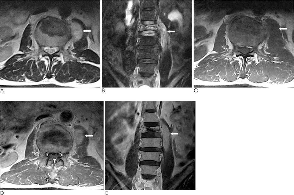
Fig. 1 A, B. Axial (A) and coronal (B) fat saturated T2-weighted turbo spin-echo (TR/TE 4230/104 ms) MR images performed on the fifth post-trauma day show an L3 compression fracture and a left psoas mass (arrows). The mass is of high signal intensity with no peripheral hypointense rim on T2-weighted turbo spin-echo image C. Axial T1-weighted spin-echo (TR/TE, 727/13 ms) MR image shows an isointense mass (arrow) relative to the muscle. D, E. Gadolinium-enhanced axial (D) and coronal (E) T1-weighted spin-echo MR images show diffuse homogenous enhancement of the mass without a central necrotic portion (arrows).

Fig. 2 A, B. Unenhanced axial (A) and reformatted sagittal (B) CT scan performed on the third post-trauma week demonstrate a well-defined, ossified peripheral rim and a central non-ossified area of low density in the lesion (arrows).
Reference
-
1. Kransdorf MJ, Meis JM, Jelinek JS. Myositis ossificans: MR appearence with radiologic-pathologic correlation. AJR Am J Roentgenol. 1991; 157:1243–1248.2. Sirvanci M, Ganiyusufoglu AK, Karaman K, Tezer M, Hamzaoglu A. Myositis ossificans of psoas muscle: magnetic resonance imaging findings. Acta Radiol. 2004; 45:523–525.3. Parikh J, Hyare H, Saifuddin A. The imaging features of post-traumatic myositis ossificans, with emphasis on MRI. Clin Radiol. 2002; 57:1058–1066.4. Ehara S, Shiraishi H, Abe M, Mizutani H. Reactive heterotopic ossification. Its patterns on MRI. Clin Imaging. 1998; 22:292–296.5. Kransdorf MJ, Meis JM. From the achives of the AFIP. Extraskeletal osseous and cartilaginous tumors of the extremities. Radiographics. 1993; 13:853–884.6. Saussez S, Blaivie C, Lemort M, Chantrain G. Non-traumatic myositis ossificans in the paraspinal muscles. Eur Arch Otorhinolaryngol. 2006; 263:331–335.7. Kim SW, Choi JH. Myositis ossificans in psoas muscle after lumbar spine fracture. Spine (Phila Pa 1976). 2009; 34:367–370.
