J Korean Soc Radiol.
2010 Jul;63(1):33-39. 10.3348/jksr.2010.63.1.33.
The Location and Size of Pulmonary Embolism in Antineoplastic Chemotherapy Patients
- Affiliations
-
- 1Department of Radiology, Wonju Christian Hospital, Yonsei University Wonju College of Medicine, Korea. wckwon@yonsei.ac.kr
- 2Department of Internal Medicine, Wonju Christian Hospital, Yonsei University Wonju College of Medicine, Korea.
- 3Department of Prevent Medicine, Wonju Christian Hospital, Yonsei University Wonju College of Medicine, Korea.
- KMID: 2208883
- DOI: http://doi.org/10.3348/jksr.2010.63.1.33
Abstract
- PURPOSE
To retrospectively evaluate the prevalent location and size of pulmonary embolism (PE) in anti-neoplastic chemotherapy patients by multidetector row CT (MDCT).
MATERIALS AND METHODS
This study was conducted on 101 patients that were positively diagnosed with PE by CT. Among these patients, 23 had received or were undergoing chemotherapy. The location and the mean size of the largest PE were compared between anti-neoplastic chemotherapy patients and non-cancer patients using the Chisquare test and paired t-test, respectively. We also used a multiple linear regression to assess the risk posed by the other risk factors of PE.
RESULTS
The most prevalent location of PE in patients on anti-neoplastic chemotherapy was in the lobar or segmental pulmonary arteries and was not significantly different from non-cancer patients. The size of the PE was smaller in patients on anti-neoplastic chemotherapy (1.14 mL [standard error = 0.29]) compared to non-cancer patients. (2.14 mL [standard error = 0.40]) (p < 0.05).
CONCLUSION
The size of PE is smaller in anti-neoplastic chemotherapy patients than in non-cancer patients.
MeSH Terms
Figure
-

Fig. 1 Axial chest CT scan shows the measurement of the embolus area using VoxelPlus. The measured area of the emboli is summed up to calculate the volume.
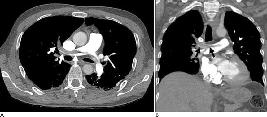
Fig. 2 63-year-old man without underlying disease. A, B. Axial and coronal CT scan show a large PE (arrow) in the main pulmonary artery.

Fig. 3 64-year-old man with lung cancer. A, B. Axial and coronal CT scan show a small PE in the laterobasal segmental artery of the left lower lobe (arrow).
Reference
-
1. de Monye W, van Strijen MJ, Huisman MV, Kieft GJ, Pattynama PM. Suspected pulmonary embolism: prevalence and anatomic distribution in 487 consecutive patients. Advances in New Technologies Evaluating the Localisation of Pulmonary Embolism (ANTELOPE) Group. Radiology. 2000; 215:184–188.2. Falanga A, Donati MB. Pathogenesis of thrombosis in patients with malignancy. Int J Hematol. 2001; 73:137–144.3. Schoepf UJ, Holzknecht N, Helmberger TK, Crispin A, Hong C, Becker CR, et al. Subsegmental pulmonary emboli: improved detection with thin-collimation multi-detector row spiral CT. Radiology. 2002; 222:483–490.4. Oser RF, Zuckerman DA, Gutierrez FR, Brink JA. Anatomic distribution of pulmonary emboli at pulmonary angiography: implications for cross-sectional imaging. Radiology. 1996; 199:31–35.5. Morgenthaler TI, Ryu JH. Clinical characteristics of fatal pulmonary embolism in a referral hospital. Mayo Clin Proc. 1995; 70:417–424.6. Gladish GW, Choe DH, Marom EM, Sabloff BS, Broemeling LD, Munden RF. Incidental pulmonary emboli in oncology patients: prevalence, CT evaluation, and natural history. Radiology. 2006; 240:246–255.7. Qanadli SD, Hajjam ME, Mesurolle B, Barre O, Bruckert F, Joseph T, et al. Pulmonary embolism detection: prospective evaluation of dual-section helical CT versus selective pulmonary arteriography in 157 patients. Radiology. 2000; 217:447–455.
- Full Text Links
-
- Actions
-
Cited
- CITED
-
- Close
- Share
-
- Similar articles
-
- Pulmonary Embolism and Pulmonary Infarction
- Acute Pulmonary Embolism
- Pulmonary Embolism Detected after Induction of the General Anesthesia: A Case Report
- A Case of Fatal Pulmonary Embolism after Arthroscopic Partial Meniscectomy
- A Case of Impending Paradoxical Embolus in a Patient with Acute Pulmonary Embolism
