Intra-Articular Nodular Fasciitis of the Knee: A Case Report of MRI Findings
- Affiliations
-
- 1Department of Radiology, Yeungnam University College of Medicine, Daegu, Korea. khcho@med.yu.ac.kr
- 2Department of Pathology, Yeungnam University College of Medicine, Daegu, Korea.
- KMID: 2208795
- DOI: http://doi.org/10.3348/jksr.2014.71.5.254
Abstract
- Nodular fasciitis, a benign soft tissue tumor consisting of myofibroblastic proliferation, is commonly located in the subcutaneous or inter- or intra-muscular layer of extremites. Intra-articular nodular fasciitis is extremely rare. We report a case of MRI findings of a nodular fasciitis in the knee of a 13-year-old boy which was removed by arthroscopic surgery and pathologically confirmed.
Figure
-
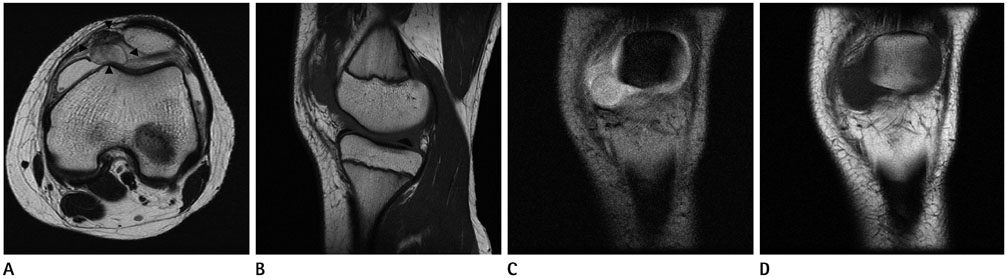
Fig. 1 Magnetic resonance imaging in a 13-year-old man with intra-articular nodular fasciitis of left knee. A. Fast spin echo axial T2-weighted image shows a well defined mass (arrowheads) with a increased signal intensity compared to normal muscle. Area of low signal intensity suggesting collagen-rich stroma is seen in ventral aspect of the mass. This mass is located in the joint capsule of inferomedial aspect of patella. Joint effusion is also noted. B. Sagittal T1-weighted image reveals iso- to slightly high signal intensity to the normal muscle. C. Gradient coronal T2-weighted image demonstrates well defined homogeneous high signal intensity. D. Coronal T1-weighted image also shows well defined iso- to slightly high signal intensity of the mass.
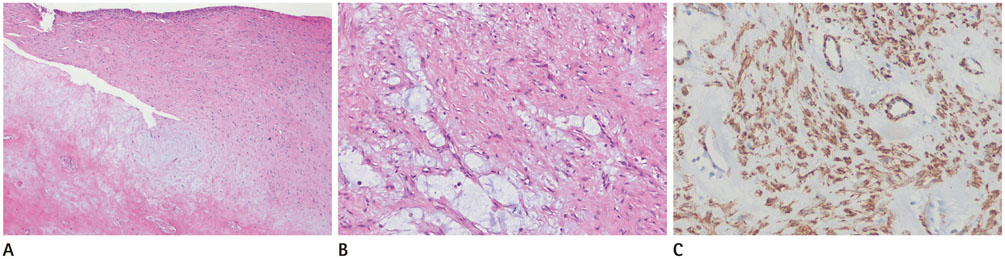
Fig. 2 Pathologic images of the intra-articular nodular fasciitis. A. Microphotograph shows myofibroblastic cell proliferation in the subsynovial layer (H&E; × 40). B. Mixed collagenous and myxoid stroma is seen (H&E; × 100). C. Immunohistochemical staining shows a positive reaction with a-smooth-muscle actin (× 200).
Reference
-
1. Sheldon PJ, Forrester DM, Learch TJ. Imaging of intraarticular masses. Radiographics. 2005; 25:105–119.2. Weiss Sw, Goldblum Jr. Enzinger and Weiss's soft tissue tumors. 5th ed. St. Louis: Mosby;2008. p. 190–194.3. Hornick JL, Fletcher CD. Intraarticular nodular fasciitis--a rare lesion: clinicopathologic analysis of a series. Am J Surg Pathol. 2006; 30:237–241.4. Lädermann A, Kindynis P, Taylor S, Ceroni D, Hoffmeyer P, Kaelin A, et al. Articular nodular fasciitis in the glenohumeral joint. Skeletal Radiol. 2008; 37:663–666.5. Gans I, Morrison MJ 3rd, Chikwava KR, Wells L. Intra-articular nodular fasciitis of the knee in a pediatric patient. Orthopedics. 2014; 37:e313–e316.6. Matsuzaki T, Akisue T, Kishimoto K, Kishimoto S, Imabori M, Hara H, et al. Intra-articular nodular fasciitis of the knee: a rare cause of recurrent hemarthrosis. Rheumatol Int. 2012; 32:1691–1694.7. Hagino T, Ochiai S, Sato E, Watanabe Y, Senga S, Kondo T, et al. Intraarticular nodular fasciitis causing limitation of knee extension: a case report. Knee. 2010; 17:424–427.8. Coyle J, White LM, Dickson B, Ferguson P, Wunder J, Naraghi A. MRI characteristics of nodular fasciitis of the musculoskeletal system. Skeletal Radiol. 2013; 42:975–982.9. Leung LY, Shu SJ, Chan AC, Chan MK, Chan CH. Nodular fasciitis: MRI appearance and literature review. Skeletal Radiol. 2002; 31:9–13.10. Botez P, Sirbu PD, Grierosu C, Mihailescu D, Savin L, Scarlat MM. Adult multifocal pigmented villonodular synovitis--clinical review. Int Orthop. 2013; 37:729–733.
