A Rare Radiological Manifestation of Disseminated Tuberculous Spondylitisin Acquired Immune Deficiency Syndrome Patient: A Case Report
- Affiliations
-
- 1Department of Radiology, Dongguk University Ilsan Hospital, Dongguk University School of Medicine, Goyang, Korea. koojb@dumc.or.kr
- KMID: 2208787
- DOI: http://doi.org/10.3348/jksr.2016.74.4.273
Abstract
- The spine is the most common site of skeletal involvement in tuberculosis. The radiologic features are reportedly characterized by destruction of the vertebral body, subligamentous extension or subchondral penetration, frequent paravertebral abscess formation and late involvement of the disk space. We experienced a case of a 25-year-old male who was a human immunodeficiency virus carrier without antiretroviral therapy. Incidental findings on abdominal computed tomography included multiple well-demarcated and ovoid osteolytic lesions with hyperdense rims disseminated in the thoracic, lumbar, and sacrum vertebrae, as well as in both ilii. On the lumbar spine magnetic resonance imaging, multiple small round lesions of isointense signal intensity with peripheral hyperintense rims were found on both T1- and T2-weighted imaging. The lesions had peripheral rim enhancement on gadolinium-enhanced T1-weighted imaging. Based on our experience, this rare image finding is one of the manifestations of disseminated tuberculosis.
MeSH Terms
Figure
-
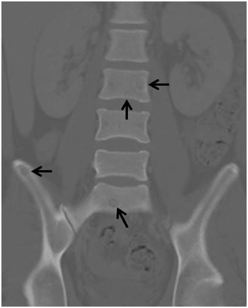
Fig. 1 Coronal-reformatted abdominal CT image shows multiple well-defined, ovoid, osteolytic lesions in the L3 vertebra, sacrum and right ilium (arrows).

Fig. 2 T1-weighted, T2-weighted, and gadolinium-enhanced T1-weighted fat-saturated sagittal lubar spine MRI images. A, B. T1-weighted (A) and T2-weighted (B) sagittal lumbar spine MRI images reveal well-defined, isointense, ovoid lesions on the lumbar spine. Thin, rim-like hyperintensity was also noted on peripheral portion of the lesions. No vertebral destruction, disk involvement or paraspinal abscess were noted on the images. C. Gadolinium-enhanced, T1 fat-saturated sagittal imaging shows peripheral enhancement of the ovoid lesions on the lumbar spine and sacrum, accentuating the target-like appearance of the lesions.

Fig. 3 Pathologic features of spinal tuberculosis. A. The biopsied spinal tissue showed caseation necrosis, a small granulomatous focus (empty arrows) with multinucleated giant cells (black arrows) (hematoxylin-eosin staining, × 400). B. Ziehl-Nielsen staining revealed acid-fast bacilli in the caseation necrosis area (Ziehl-Nielsen staining, × 400).
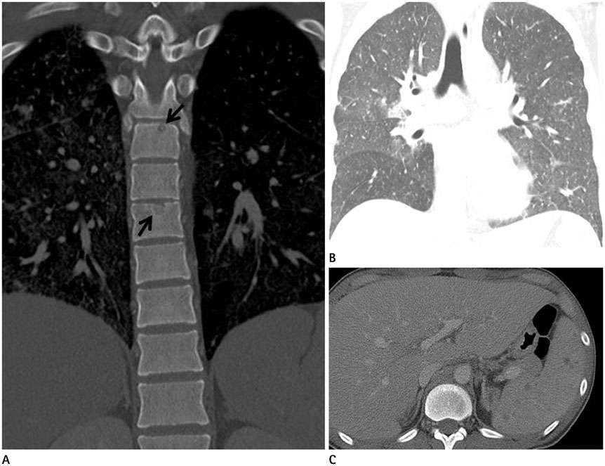
Fig. 4 Miliary tuberculosis on follow-up chest CT after 2 months. A. Coronal-reformatted chest CT image shows multiple well-defined, ovoid, osteolytic lesions in thoracic vertebrae (arrows). B. Coronal-reformatted chest CT image shows numerous random distributed miliary nodules. C. Axial CT image shows several ill-demarcated round low density lesions in the spleen.
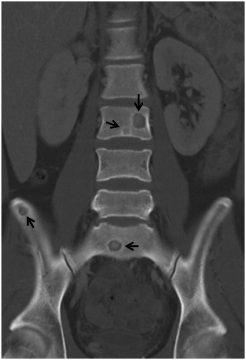
Fig. 5 Coronal-reformatted abdominal CT image shows multiple well-defined, ovoid, osteolytic lesions in the L3 vertebra, sacrum and right ilium (arrows).
Reference
-
1. Tehranzadeh J, Ter-Oganesyan RR, Steinbach LS. Musculoskeletal disorders associated with HIV infection and AIDS. Part II: non-infectious musculoskeletal conditions. Skeletal Radiol. 2004; 33:311–320.2. Restrepo CS, Lemos DF, Gordillo H, Odero R, Varghese T, Tiemann W, et al. Imaging findings in musculoskeletal complications of AIDS. Radiographics. 2004; 24:1029–1049.3. Moore SL, Rafii M. Imaging of musculoskeletal and spinal tuberculosis. Radiol Clin North Am. 2001; 39:329–342.4. Shanley DJ. Tuberculosis of the spine: imaging features. AJR Am J Roentgenol. 1995; 164:659–664.5. Restrepo CS, Martínez S, Lemos JA, Carrillo JA, Lemos DF, Ojeda P, et al. Imaging manifestations of Kaposi sarcoma. Radiographics. 2006; 26:1169–1185.6. Ahmadi J, Bajaj A, Destian S, Segall HD, Zee CS. Spinal tuberculosis: atypical observations at MR imaging. Radiology. 1993; 189:489–493.
- Full Text Links
-
- Actions
-
Cited
- CITED
-
- Close
- Share
-
- Similar articles
-
- Tuberculous Epididymo-orchitis in Acquired Immune Deficiency Syndrome Patients
- Molluscum Contagiosum as a Skin Manifestation of Immune Reconstitution Inflammatory Syndrome in an AIDS Patient Who Is Receiving HAART
- A Case of Cytomegalovirus Lumbosacral Polyradiculopathy in Acquired Immune Deficiency Syndrome
- Acquired immune deficiency syndrome: report of an autopsy case
- Acquired C1 inhibitor deficiency associated with SLE
